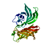
- PDB-3uwt: Crystal structure of a RNA binding domain of poly-U binding splic... -
+
Open data
ID or keywords:
Loading...
-
Basic information
Entry
Database: PDB / ID: 3uwt
Title
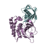
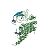
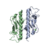
Crystal structure of a RNA binding domain of poly-U binding splicing factor 60KDa (PUF60) from Homo sapiens at 2.50 A resolution
Components
Poly(U)-binding-splicing factor PUF60
Keywords
RNA BINDING PROTEIN / RNA recognition motive / RRM / RNA binding domain / splicing / Structural Genomics / Joint Center for Structural Genomics / JCSG / Protein Structure Initiative / PSI-BIOLOGY / Partnership for T-Cell Biology / TCELL
Function / homology
Function and homology information
mRNA splice site recognition / alternative mRNA splicing, via spliceosome / regulation of alternative mRNA splicing, via spliceosome / mRNA Splicing - Major Pathway / cell junction / cadherin binding / ribonucleoprotein complex / apoptotic process / DNA binding / RNA binding ...mRNA splice site recognition / alternative mRNA splicing, via spliceosome / regulation of alternative mRNA splicing, via spliceosome / mRNA Splicing - Major Pathway / cell junction / cadherin binding / ribonucleoprotein complex / apoptotic process / DNA binding / RNA binding / nucleoplasm / identical protein binding Similarity search - Function
Mass: 18.015 Da / Num. of mol.: 27 / Source method: isolated from a natural source / Formula: H2O
Has protein modification
Y
Sequence details
THIS CONSTRUCT (RESIDUES 118-316) WAS EXPRESSED WITH A PURIFICATION TAG MGSDKIHHHHHHENLYFQG. THE ...THIS CONSTRUCT (RESIDUES 118-316) WAS EXPRESSED WITH A PURIFICATION TAG MGSDKIHHHHHHENLYFQG. THE TAG WAS REMOVED WITH TEV PROTEASE LEAVING ONLY A GLYCINE (0) FOLLOWED BY THE TARGET SEQUENCE. RESIDUE NUMBERING IS BASED ON ISOFORM 1 OF UNIPROTKB Q9UHX1.
-
Experimental details
-
Experiment
Experiment
Method: X-RAY DIFFRACTION / Number of used crystals: 1
-
Sample preparation
Crystal
Density Matthews: 2.14 Å3/Da / Density % sol: 42.62 %
Crystal grow
Temperature: 293 K / Method: vapor diffusion, sitting drop / pH: 9.5 Details: 0.2M sodium chloride, 1.26M ammonium sulfate, 0.1M CHES pH 9.5, NANODROP, VAPOR DIFFUSION, SITTING DROP, temperature 293K
Resolution: 2.5→29.798 Å / Num. all: 6937 / Num. obs: 6937 / % possible obs: 99.8 % / Redundancy: 6.5 % / Biso Wilson estimate: 68.061 Å2 / Rsym value: 0.094 / Net I/σ(I): 11.4
Reflection shell
Diffraction-ID: 1
Resolution (Å)
Redundancy (%)
Mean I/σ(I) obs
Num. measured all
Num. unique all
Rsym value
% possible all
Rmerge(I) obs
2.5-2.57
6.9
1.6
3532
514
1.377
99.9
2.57-2.64
6.8
0.7
3254
478
1.003
99.6
0.014
2.64-2.71
6.7
1
3205
475
0.724
99.7
0.014
2.71-2.8
6.8
1.3
3200
473
0.595
99.8
0.014
2.8-2.89
6.8
1.9
2982
439
0.406
99.8
0.014
2.89-2.99
6.7
2.5
2871
430
0.304
99.8
0.014
2.99-3.1
6.8
3.2
2887
426
0.237
100
0.014
3.1-3.23
6.7
4.6
2691
402
0.163
99.8
0.014
3.23-3.37
6.6
5.8
2613
395
0.13
99.8
0.014
3.37-3.54
6.6
7.4
2502
377
0.098
99.8
0.014
3.54-3.73
6.3
5.8
2221
353
0.118
99.9
0.014
3.73-3.95
6.4
9.6
2222
348
0.071
99.9
0.014
3.95-4.23
6.4
11
2015
314
0.061
100
0.014
4.23-4.56
6.4
12.9
1919
301
0.05
99.9
0.014
4.56-5
6.1
12
1728
281
0.051
99.8
0.014
5-5.59
5.9
10.6
1492
251
0.055
99.9
0.014
5.59-6.46
5.6
9.1
1310
234
0.071
99.7
0.014
6.46-7.91
6.1
8.8
1209
197
0.07
99.9
0.014
7.91-11.18
6.2
12.1
970
157
0.052
99.7
0.014
11.18-29.798
5.5
13.9
508
92
0.045
94.7
0.014
-
Phasing
Phasing
Method: MAD
-
Processing
Software
Name
Version
Classification
NB
MolProbity
3beta29
modelbuilding
PDB_EXTRACT
3.1
dataextraction
SHELX
phasing
SHARP
phasing
SCALA
3.3.20
datascaling
BUSTER-TNT
2.10.0
refinement
MOSFLM
datareduction
SHELXD
phasing
BUSTER
2.10.0
refinement
Refinement
Method to determine structure: MAD / Resolution: 2.5→29.798 Å / Cor.coef. Fo:Fc: 0.9446 / Cor.coef. Fo:Fc free: 0.9143 / Occupancy max: 1 / Occupancy min: 0.5 / Cross valid method: THROUGHOUT / σ(F): 0 Details: 1. A MET-INHIBITION PROTOCOL WAS USED FOR SELENOMETHIONINE INCORPORATION DURING PROTEIN EXPRESSION. THE OCCUPANCY OF THE SE ATOMS IN THE MSE RESIDUES WAS REDUCED TO 0.75 FOR THE REDUCED ...Details: 1. A MET-INHIBITION PROTOCOL WAS USED FOR SELENOMETHIONINE INCORPORATION DURING PROTEIN EXPRESSION. THE OCCUPANCY OF THE SE ATOMS IN THE MSE RESIDUES WAS REDUCED TO 0.75 FOR THE REDUCED SCATTERING POWER DUE TO PARTIAL S-MET INCORPORATION. 2. ATOM RECORD CONTAINS SUM OF TLS AND RESIDUAL B FACTORS. ANISOU RECORD CONTAINS SUM OF TLS AND RESIDUAL U FACTORS. 3. CHLORIDE (CL) FROM THE CRYSTALLIZATION SOLUTION HAS BEEN MODELED IN THE SOLVENT STRUCTURE. 4. THE REFINEMENT WAS RESTRAINED WITH THE MAD PHASES. 5. RAMACHANDRAN OUTLIERS AT RESIDUES 150 AND 290 ARE SUPPORTED BY ELECTRON DENSITY.
In the structure databanks used in Yorodumi, some data are registered as the other names, "COVID-19 virus" and "2019-nCoV". Here are the details of the virus and the list of structure data.
Jan 31, 2019. EMDB accession codes are about to change! (news from PDBe EMDB page)
EMDB accession codes are about to change! (news from PDBe EMDB page)
The allocation of 4 digits for EMDB accession codes will soon come to an end. Whilst these codes will remain in use, new EMDB accession codes will include an additional digit and will expand incrementally as the available range of codes is exhausted. The current 4-digit format prefixed with “EMD-” (i.e. EMD-XXXX) will advance to a 5-digit format (i.e. EMD-XXXXX), and so on. It is currently estimated that the 4-digit codes will be depleted around Spring 2019, at which point the 5-digit format will come into force.
The EM Navigator/Yorodumi systems omit the EMD- prefix.
Related info.:Q: What is EMD? / ID/Accession-code notation in Yorodumi/EM Navigator
Yorodumi is a browser for structure data from EMDB, PDB, SASBDB, etc.
This page is also the successor to EM Navigator detail page, and also detail information page/front-end page for Omokage search.
The word "yorodu" (or yorozu) is an old Japanese word meaning "ten thousand". "mi" (miru) is to see.
Related info.:EMDB / PDB / SASBDB / Comparison of 3 databanks / Yorodumi Search / Aug 31, 2016. New EM Navigator & Yorodumi / Yorodumi Papers / Jmol/JSmol / Function and homology information / Changes in new EM Navigator and Yorodumi
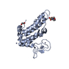
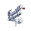
 Movie
Movie Controller

 Yorodumi
 Open data
 Basic information
 Components
 Keywords
 Function and homology information
 Homo sapiens (human)
 X-RAY DIFFRACTION /
  Authors
 Citation
 Structure visualization
 Downloads & links
 Other downloads
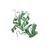


 PDBj
PDBj


 Assembly
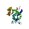


 Mass: 35.453 Da / Num. of mol.: 1 / Source method: obtained synthetically / Formula: Cl
 Mass: 18.015 Da / Num. of mol.: 27 / Source method: isolated from a natural source / Formula: H2O
 Sample preparation
 / Beamline: 8.2.2 / Wavelength: 0.9537,0.9796,0.9793
 Processing