 Movie
Movie Controller

[English] 日本語
 Yorodumi
- PDB-5kwg: Crystal structure of extracellular domain of HER2 in complex with... -
+ Open data

- Basic information
| Entry | Database: PDB / ID: 5kwg | |||||||||
|---|---|---|---|---|---|---|---|---|---|---|
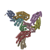
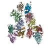
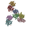
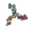
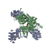
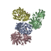
| Title | Crystal structure of extracellular domain of HER2 in complex with Fcab H10-03-6 | |||||||||
 Components Components |
| |||||||||
 Keywords Keywords | IMMUNE SYSTEM / antibody engineering / immunoglobulin G1 / Fc fragment / glycosylations / CH3 domain / Fcab / human epidermal growth factor receptor 2 / HER2/neu / erbB-2 / cell surface receptor | |||||||||
| Function / homology |  Function and homology information Function and homology informationnegative regulation of immature T cell proliferation in thymus / ERBB3:ERBB2 complex / ERBB2-ERBB4 signaling pathway / immature T cell proliferation in thymus / GRB7 events in ERBB2 signaling / RNA polymerase I core binding / semaphorin receptor complex / Developmental Lineage of Mammary Stem Cells / ErbB-3 class receptor binding / complement-dependent cytotoxicity ...negative regulation of immature T cell proliferation in thymus / ERBB3:ERBB2 complex / ERBB2-ERBB4 signaling pathway / immature T cell proliferation in thymus / GRB7 events in ERBB2 signaling / RNA polymerase I core binding / semaphorin receptor complex / Developmental Lineage of Mammary Stem Cells / ErbB-3 class receptor binding / complement-dependent cytotoxicity / antibody-dependent cellular cytotoxicity / Sema4D induced cell migration and growth-cone collapse / Fc-gamma receptor I complex binding / motor neuron axon guidance / regulation of microtubule-based process / immunoglobulin complex, circulating / Classical antibody-mediated complement activation / immunoglobulin receptor binding / Initial triggering of complement / IgG immunoglobulin complex / PLCG1 events in ERBB2 signaling / enzyme-linked receptor protein signaling pathway / ERBB2 Activates PTK6 Signaling / ERBB2-EGFR signaling pathway / ERBB2-ERBB3 signaling pathway / neurotransmitter receptor localization to postsynaptic specialization membrane / Drug-mediated inhibition of ERBB2 signaling / Resistance of ERBB2 KD mutants to trastuzumab / Resistance of ERBB2 KD mutants to sapitinib / Resistance of ERBB2 KD mutants to tesevatinib / Resistance of ERBB2 KD mutants to neratinib / Resistance of ERBB2 KD mutants to osimertinib / Resistance of ERBB2 KD mutants to afatinib / Resistance of ERBB2 KD mutants to AEE788 / Resistance of ERBB2 KD mutants to lapatinib / Drug resistance in ERBB2 TMD/JMD mutants / positive regulation of MAP kinase activity / neuromuscular junction development / positive regulation of Rho protein signal transduction / positive regulation of transcription by RNA polymerase I / ERBB2 Regulates Cell Motility / Developmental Lineage of Mammary Gland Myoepithelial Cells / oligodendrocyte differentiation / semaphorin-plexin signaling pathway / FCGR activation / PI3K events in ERBB2 signaling / complement activation, classical pathway / regulation of angiogenesis / Role of phospholipids in phagocytosis / positive regulation of protein targeting to membrane / regulation of ERK1 and ERK2 cascade / Schwann cell development / antigen binding / coreceptor activity / Signaling by ERBB2 / TFAP2 (AP-2) family regulates transcription of growth factors and their receptors / peptidyl-tyrosine phosphorylation / myelination / transmembrane receptor protein tyrosine kinase activity / GRB2 events in ERBB2 signaling / FCGR3A-mediated IL10 synthesis / positive regulation of cell adhesion / cell surface receptor protein tyrosine kinase signaling pathway / Regulation of Complement cascade / SHC1 events in ERBB2 signaling / basal plasma membrane / cellular response to epidermal growth factor stimulus / Constitutive Signaling by Overexpressed ERBB2 / positive regulation of epithelial cell proliferation / Downregulation of ERBB2:ERBB3 signaling / B cell receptor signaling pathway / positive regulation of translation / FCGR3A-mediated phagocytosis / neuromuscular junction / wound healing / phosphatidylinositol 3-kinase/protein kinase B signal transduction / Signaling by ERBB2 TMD/JMD mutants / receptor protein-tyrosine kinase / Signaling by ERBB2 ECD mutants / Signaling by ERBB2 KD Mutants / Regulation of actin dynamics for phagocytic cup formation / receptor tyrosine kinase binding / cellular response to growth factor stimulus / epidermal growth factor receptor signaling pathway / ruffle membrane / Downregulation of ERBB2 signaling / Constitutive Signaling by Aberrant PI3K in Cancer / neuron differentiation / transmembrane signaling receptor activity / PIP3 activates AKT signaling / myelin sheath / antibacterial humoral response / heart development / PI5P, PP2A and IER3 Regulate PI3K/AKT Signaling / positive regulation of cell growth / RAF/MAP kinase cascade / protein tyrosine kinase activity / Interleukin-4 and Interleukin-13 signaling / presynaptic membrane / blood microparticle Similarity search - Function | |||||||||
| Biological species |  Homo sapiens (human) Homo sapiens (human) | |||||||||
| Method |  X-RAY DIFFRACTION / SYNCHROTRON / MOLECULAR REPLACEMENT / Resolution: 4.3 Å X-RAY DIFFRACTION / SYNCHROTRON / MOLECULAR REPLACEMENT / Resolution: 4.3 Å | |||||||||
 Authors Authors | Humm, A. / Lobner, E. / Goritzer, K. / Mlynek, G. / Obinger, C. / Djinovic-Carugo, K. | |||||||||
| Funding support |  Austria, 2items Austria, 2items
| |||||||||
 Citation Citation | Journal: Structure / Year: 2017 Title: Fcab-HER2 Interaction: a Menage a Trois. Lessons from X-Ray and Solution Studies. Authors: Lobner, E. / Humm, A.S. / Goritzer, K. / Mlynek, G. / Puchinger, M.G. / Hasenhindl, C. / Ruker, F. / Traxlmayr, M.W. / Djinovic-Carugo, K. / Obinger, C. | |||||||||
| History |
|
- Structure visualization
| Structure viewer | Molecule: MolmilJmol/JSmol |
|---|
- Downloads & links
-Download
| PDBx/mmCIF format | 5kwg.cif.gz | 439.5 KB | Display | PDBx/mmCIF format |
|---|---|---|---|---|
| PDB format | pdb5kwg.ent.gz | 366.8 KB | Display | PDB format |
| PDBx/mmJSON format | 5kwg.json.gz | Tree view | PDBx/mmJSON format | |
| Others |  Other downloads Other downloads |
-Validation report
| Arichive directory | https://data.pdbj.org/pub/pdb/validation_reports/kw/5kwgftp://data.pdbj.org/pub/pdb/validation_reports/kw/5kwg | HTTPS FTP |
|---|
-Related structure data
| Related structure data |  5jihC  5jiiC 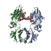 5jikC 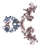 5k33SC S: Starting model for refinement C: citing same article ( |
|---|---|
| Similar structure data |


-Links
 PDBj
PDBj


- Assembly
| Deposited unit | 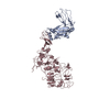
| ||||||||
|---|---|---|---|---|---|---|---|---|---|
| 1 | 
| ||||||||
| Unit cell |
|
-Components
| #1: Protein | Mass: 69507.891 Da / Num. of mol.: 1 Source method: isolated from a genetically manipulated source Source: (gene. exp.) Homo sapiens (human) / Gene: ERBB2, HER2, MLN19, NEU, NGL / Cell line (production host): CHO Lec1 / Organ (production host): Ovary / Production host:   Cricetulus griseus (Chinese hamster) Cricetulus griseus (Chinese hamster)References: UniProt: P04626, receptor protein-tyrosine kinase |
|---|---|
| #2: Protein | Mass: 25770.238 Da / Num. of mol.: 1 Source method: isolated from a genetically manipulated source Source: (gene. exp.) Homo sapiens (human) / Gene: IGHG1 / Plasmid: Plasmid / Cell line (production host): HEK 293-6E / Organ (production host): Kidney / Production host: Homo sapiens (human) / References: UniProt: P01857 |
| Has protein modification | Y |
-Experimental details
-Experiment
| Experiment | Method: X-RAY DIFFRACTION / Number of used crystals: 1 |
|---|
- Sample preparation
| Crystal | Density Matthews: 3.26 Å3/Da / Density % sol: 62.3 % |
|---|---|
| Crystal grow | Temperature: 295 K / Method: vapor diffusion, sitting drop / pH: 6 / Details: 50 mM Citric acid, 18% (w/v) PEG 3350 |
-Data collection
| Diffraction | Mean temperature: 100 K |
|---|---|
| Diffraction source | Source: SYNCHROTRON / Site: ESRF  / Beamline: MASSIF-3 / Wavelength: 0.9677 Å / Beamline: MASSIF-3 / Wavelength: 0.9677 Å |
| Detector | Type: DECTRIS PILATUS3 2M / Detector: PIXEL / Date: Nov 5, 2015 / Details: CRL |
| Radiation | Monochromator: C(110) / Protocol: SINGLE WAVELENGTH / Monochromatic (M) / Laue (L): M / Scattering type: x-ray |
| Radiation wavelength | Wavelength: 0.9677 Å / Relative weight: 1 |
| Reflection | Resolution: 4.3→47.532 Å / Num. all: 55512 / Num. obs: 8755 / % possible obs: 100 % / Redundancy: 6.3 % / Biso Wilson estimate: 174.26 Å2 / CC1/2: 0.998 / Rmerge(I) obs: 0.1898 / Net I/σ(I): 6.48 |
| Reflection shell | Resolution: 4.3→4.454 Å / Redundancy: 6 % / Rmerge(I) obs: 1.55 / Mean I/σ(I) obs: 0.99 / % possible all: 100 |
- Processing
| Software |
| |||||||||||||||||||||||||||||||||||||||||||||||||||||||||||||||||||||||||||||||||||||||||||||||||||||||||||||||||||||||||||||||||||||||||||||||||||||||||||||||||||||||||||||||
|---|---|---|---|---|---|---|---|---|---|---|---|---|---|---|---|---|---|---|---|---|---|---|---|---|---|---|---|---|---|---|---|---|---|---|---|---|---|---|---|---|---|---|---|---|---|---|---|---|---|---|---|---|---|---|---|---|---|---|---|---|---|---|---|---|---|---|---|---|---|---|---|---|---|---|---|---|---|---|---|---|---|---|---|---|---|---|---|---|---|---|---|---|---|---|---|---|---|---|---|---|---|---|---|---|---|---|---|---|---|---|---|---|---|---|---|---|---|---|---|---|---|---|---|---|---|---|---|---|---|---|---|---|---|---|---|---|---|---|---|---|---|---|---|---|---|---|---|---|---|---|---|---|---|---|---|---|---|---|---|---|---|---|---|---|---|---|---|---|---|---|---|---|---|---|---|---|
| Refinement | Method to determine structure: MOLECULAR REPLACEMENT Starting model: 5K33 Resolution: 4.3→47.532 Å / SU ML: 0.87 / Cross valid method: FREE R-VALUE / σ(F): 1.26 / Phase error: 41.45 / Stereochemistry target values: ML
| |||||||||||||||||||||||||||||||||||||||||||||||||||||||||||||||||||||||||||||||||||||||||||||||||||||||||||||||||||||||||||||||||||||||||||||||||||||||||||||||||||||||||||||||
| Solvent computation | Shrinkage radii: 0.9 Å / VDW probe radii: 1.11 Å / Solvent model: FLAT BULK SOLVENT MODEL | |||||||||||||||||||||||||||||||||||||||||||||||||||||||||||||||||||||||||||||||||||||||||||||||||||||||||||||||||||||||||||||||||||||||||||||||||||||||||||||||||||||||||||||||
| Refinement step | Cycle: LAST / Resolution: 4.3→47.532 Å
| |||||||||||||||||||||||||||||||||||||||||||||||||||||||||||||||||||||||||||||||||||||||||||||||||||||||||||||||||||||||||||||||||||||||||||||||||||||||||||||||||||||||||||||||
| Refine LS restraints |
| |||||||||||||||||||||||||||||||||||||||||||||||||||||||||||||||||||||||||||||||||||||||||||||||||||||||||||||||||||||||||||||||||||||||||||||||||||||||||||||||||||||||||||||||
| LS refinement shell |
| |||||||||||||||||||||||||||||||||||||||||||||||||||||||||||||||||||||||||||||||||||||||||||||||||||||||||||||||||||||||||||||||||||||||||||||||||||||||||||||||||||||||||||||||
| Refinement TLS params. | Method: refined / Refine-ID: X-RAY DIFFRACTION
| |||||||||||||||||||||||||||||||||||||||||||||||||||||||||||||||||||||||||||||||||||||||||||||||||||||||||||||||||||||||||||||||||||||||||||||||||||||||||||||||||||||||||||||||
| Refinement TLS group |
|
