- PDB-5ku5: Crystal Structure of CusS Sensor Domain with Silver Bound -
+
Open data
ID or keywords:
Loading...
-
Basic information
Entry
Database: PDB / ID: 5ku5
Title
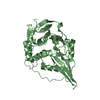
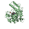
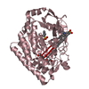
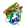
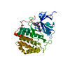
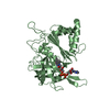
Crystal Structure of CusS Sensor Domain with Silver Bound
Components
Sensor kinase CusS
Keywords
TRANSFERASE / PDC fold / Histidine kinase / Silver binding / Metal efflux system
Function / homology
Function and homology information
cellular response to silver ion / protein histidine kinase activity / phosphorelay sensor kinase activity / histidine kinase / phosphorelay signal transduction system / cellular response to copper ion / signal transduction / ATP binding / membrane / metal ion binding / plasma membrane Similarity search - Function
: / CusS sensor domain / Heavy metal sensor kinase / : / HAMP domain / HAMP (Histidine kinases, Adenylyl cyclases, Methyl binding proteins, Phosphatases) domain / HAMP domain profile. / HAMP domain / His Kinase A (phospho-acceptor) domain / His Kinase A (phosphoacceptor) domain ...: / CusS sensor domain / Heavy metal sensor kinase / : / HAMP domain / HAMP (Histidine kinases, Adenylyl cyclases, Methyl binding proteins, Phosphatases) domain / HAMP domain profile. / HAMP domain / His Kinase A (phospho-acceptor) domain / His Kinase A (phosphoacceptor) domain / Signal transduction histidine kinase, dimerisation/phosphoacceptor domain / Signal transduction histidine kinase-related protein, C-terminal / Signal transduction histidine kinase, dimerisation/phosphoacceptor domain superfamily / Histidine kinase domain / Histidine kinase domain profile. / Histidine kinase-, DNA gyrase B-, and HSP90-like ATPase / Histidine kinase-like ATPases / Histidine kinase/HSP90-like ATPase / Histidine kinase/HSP90-like ATPase superfamily Similarity search - Domain/homology
Protocol: SINGLE WAVELENGTH / Monochromatic (M) / Laue (L): M / Scattering type: x-ray
Radiation wavelength
Wavelength: 1.036 Å / Relative weight: 1
Reflection
Resolution: 2.15→33.29 Å / Num. obs: 38599 / % possible obs: 99 % / Redundancy: 3.48 % / Net I/σ(I): 6.3
-
Processing
Software
Name
Version
Classification
REFMAC
5.8.0103
refinement
PDB_EXTRACT
3.2
dataextraction
iMOSFLM
datareduction
Aimless
datascaling
PHENIX
phasing
Refinement
Method to determine structure: SAD / Resolution: 2.15→33.29 Å / Cor.coef. Fo:Fc: 0.922 / Cor.coef. Fo:Fc free: 0.9 / SU B: 8.019 / SU ML: 0.193 / Cross valid method: THROUGHOUT / σ(F): 0 / ESU R: 0.291 / ESU R Free: 0.229 Details: HYDROGENS HAVE BEEN ADDED IN THE RIDING POSITIONS U VALUES : REFINED INDIVIDUALLY
Rfactor
Num. reflection
% reflection
Selection details
Rfree
0.2864
1910
5 %
RANDOM
Rwork
0.2497
-
-
-
obs
0.2515
36652
98.99 %
-
Solvent computation
Ion probe radii: 0.8 Å / Shrinkage radii: 0.8 Å / VDW probe radii: 1.2 Å
In the structure databanks used in Yorodumi, some data are registered as the other names, "COVID-19 virus" and "2019-nCoV". Here are the details of the virus and the list of structure data.
Jan 31, 2019. EMDB accession codes are about to change! (news from PDBe EMDB page)
EMDB accession codes are about to change! (news from PDBe EMDB page)
The allocation of 4 digits for EMDB accession codes will soon come to an end. Whilst these codes will remain in use, new EMDB accession codes will include an additional digit and will expand incrementally as the available range of codes is exhausted. The current 4-digit format prefixed with “EMD-” (i.e. EMD-XXXX) will advance to a 5-digit format (i.e. EMD-XXXXX), and so on. It is currently estimated that the 4-digit codes will be depleted around Spring 2019, at which point the 5-digit format will come into force.
The EM Navigator/Yorodumi systems omit the EMD- prefix.
Related info.:Q: What is EMD? / ID/Accession-code notation in Yorodumi/EM Navigator
Yorodumi is a browser for structure data from EMDB, PDB, SASBDB, etc.
This page is also the successor to EM Navigator detail page, and also detail information page/front-end page for Omokage search.
The word "yorodu" (or yorozu) is an old Japanese word meaning "ten thousand". "mi" (miru) is to see.
Related info.:EMDB / PDB / SASBDB / Comparison of 3 databanks / Yorodumi Search / Aug 31, 2016. New EM Navigator & Yorodumi / Yorodumi Papers / Jmol/JSmol / Function and homology information / Changes in new EM Navigator and Yorodumi
 Movie
Movie Controller


 Open data
 Basic information
 Components
 Keywords
 Function and homology information
 X-RAY DIFFRACTION /
  Authors
 United States, 1items
  Citation
 Structure visualization
 Downloads & links
 Other downloads
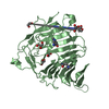


 PDBj
PDBj


 Assembly
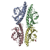

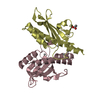
 Mass: 107.868 Da / Num. of mol.: 8 / Source method: obtained synthetically / Formula: Ag
 Mass: 59.044 Da / Num. of mol.: 1 / Source method: obtained synthetically / Formula: C2H3O2
 Mass: 18.015 Da / Num. of mol.: 207 / Source method: isolated from a natural source / Formula: H2O
 Sample preparation
 Processing