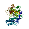
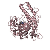
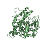
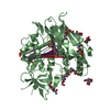
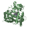
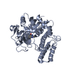
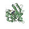
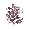
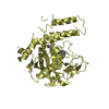
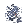
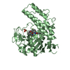
Entry Database : PDB / ID : 4hhyTitle Crystal structure of PARP catalytic domain in complex with novel inhibitors Poly [ADP-ribose] polymerase 1 Keywords / / Function / homology Function Domain/homology Component
/ / / / / / / / / / / / / / / / / / / / / / / / / / / / / / / / / / / / / / / / / / / / / / / / / / / / / / / / / / / / / / / / / / / / / / / / / / / / / / / / / / / / / / / / / / / / / / / / / / / / / / / / / / / / / / / / / / / / / / / / / / / / / / / / / / / / / / / / / / / / / / / / / / / / / / / / / / / / Biological species Homo sapiens (human)Method / / / / Resolution : 2.3637 Å Authors Liu, Q.F. / Chen, T.T. / Xu, Y.C. Journal : J.Med.Chem. / Year : 2013Title : Design, Synthesis, and Biological Evaluation of a Series of Benzo[de][1,7]naphthyridin-7(8H)-ones Bearing a Functionalized Longer Chain Appendage as Novel PARP1 Inhibitors.Authors : Ye, N. / Chen, C.H. / Chen, T. / Song, Z. / He, J.X. / Huan, X.J. / Song, S.S. / Liu, Q. / Chen, Y. / Ding, J. / Xu, Y. / Miao, Z.H. / Zhang, A. History Deposition Oct 10, 2012 Deposition site / Processing site Revision 1.0 Mar 27, 2013 Provider / Type Revision 1.1 Apr 24, 2013 Group Revision 1.2 Sep 20, 2023 Group Data collection / Database references ... Data collection / Database references / Derived calculations / Refinement description Category chem_comp_atom / chem_comp_bond ... chem_comp_atom / chem_comp_bond / database_2 / pdbx_initial_refinement_model / struct_ref_seq_dif / struct_site Item _database_2.pdbx_DOI / _database_2.pdbx_database_accession ... _database_2.pdbx_DOI / _database_2.pdbx_database_accession / _struct_ref_seq_dif.details / _struct_site.pdbx_auth_asym_id / _struct_site.pdbx_auth_comp_id / _struct_site.pdbx_auth_seq_id Revision 1.3 Oct 30, 2024 Group / Category / pdbx_modification_feature
Show all Show less
 Movie
Movie Controller

 Yorodumi
 Open data
 Basic information
 Components
 Keywords
 Function and homology information
 Homo sapiens (human)
 X-RAY DIFFRACTION /
  Authors
 Citation
 Structure visualization
 Downloads & links
 Other downloads

 PDBj
PDBj


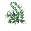
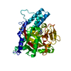
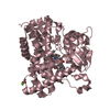
 Assembly
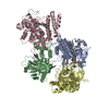

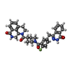
 Mass: 579.621 Da / Num. of mol.: 4 / Source method: obtained synthetically / Formula: C33H30FN5O4
 Mass: 96.063 Da / Num. of mol.: 2 / Source method: obtained synthetically / Formula: SO4
 Mass: 106.120 Da / Num. of mol.: 1 / Source method: obtained synthetically / Formula: C4H10O3
 Mass: 18.015 Da / Num. of mol.: 84 / Source method: isolated from a natural source / Formula: H2O
 Sample preparation
 / Beamline: BL17U / Wavelength: 0.979 Å
 Processing