 Movie
Movie Controller


+ Open data

- Basic information
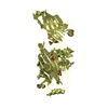
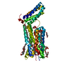
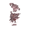
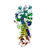
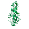
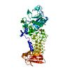
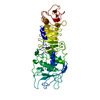
| Entry | Database: PDB / ID: 5ku1 | ||||||
|---|---|---|---|---|---|---|---|
| Title | hMiro1 EF hand and cGTPase domains in the GDP-bound state | ||||||
 Components Components | Mitochondrial Rho GTPase 1 | ||||||
 Keywords Keywords | HYDROLASE / Miro / GTPase / Parkin / Mitochondria | ||||||
| Function / homology |  Function and homology information Function and homology informationRHOT1 GTPase cycle / mitochondrial outer membrane permeabilization / cellular homeostasis / regulation of mitochondrion organization / mitochondrion transport along microtubule / mitochondrion organization / Hydrolases; Acting on acid anhydrides; Acting on GTP to facilitate cellular and subcellular movement / mitochondrial outer membrane / Ub-specific processing proteases / GTPase activity ...RHOT1 GTPase cycle / mitochondrial outer membrane permeabilization / cellular homeostasis / regulation of mitochondrion organization / mitochondrion transport along microtubule / mitochondrion organization / Hydrolases; Acting on acid anhydrides; Acting on GTP to facilitate cellular and subcellular movement / mitochondrial outer membrane / Ub-specific processing proteases / GTPase activity / calcium ion binding / GTP binding / mitochondrion / membrane Similarity search - Function | ||||||
| Biological species |  Homo sapiens (human) Homo sapiens (human) | ||||||
| Method |  X-RAY DIFFRACTION / SYNCHROTRON / MOLECULAR REPLACEMENT / Resolution: 2.501 Å X-RAY DIFFRACTION / SYNCHROTRON / MOLECULAR REPLACEMENT / Resolution: 2.501 Å | ||||||
 Authors Authors | Klosowiak, J.L. / Focia, P.J. / Rice, S.E. / Freymann, D.M. | ||||||
 Citation Citation | Journal: Sci Rep / Year: 2016 Title: Structural insights into Parkin substrate lysine targeting from minimal Miro substrates. Authors: Klosowiak, J.L. / Park, S. / Smith, K.P. / French, M.E. / Focia, P.J. / Freymann, D.M. / Rice, S.E. | ||||||
| History |
|
- Structure visualization
| Structure viewer | Molecule: MolmilJmol/JSmol |
|---|
- Downloads & links
-Download
| PDBx/mmCIF format | 5ku1.cif.gz | 98.6 KB | Display | PDBx/mmCIF format |
|---|---|---|---|---|
| PDB format | pdb5ku1.ent.gz | 72.1 KB | Display | PDB format |
| PDBx/mmJSON format | 5ku1.json.gz | Tree view | PDBx/mmJSON format | |
| Others |  Other downloads Other downloads |
-Validation report
| Summary document | 5ku1_validation.pdf.gz | 784.5 KB | Display | wwPDB validaton report |
|---|---|---|---|---|
| Full document | 5ku1_full_validation.pdf.gz | 789.1 KB | Display | |
| Data in XML | 5ku1_validation.xml.gz | 16.6 KB | Display | |
| Data in CIF | 5ku1_validation.cif.gz | 22.2 KB | Display | |
| Arichive directory | https://data.pdbj.org/pub/pdb/validation_reports/ku/5ku1ftp://data.pdbj.org/pub/pdb/validation_reports/ku/5ku1 | HTTPS FTP |
-Related structure data
| Related structure data |  5ksoC  5kspC  5ksyC  5kszC  5ktyC  5kutC  4c0lS C: citing same article ( S: Starting model for refinement |
|---|---|
| Similar structure data |

-Links
 PDBj
PDBj- Assembly
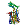
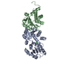
| Deposited unit | 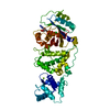
| ||||||||
|---|---|---|---|---|---|---|---|---|---|
| 1 |
| ||||||||
| Unit cell |
|
-Components
| #1: Protein | Mass: 49065.859 Da / Num. of mol.: 1 / Fragment: hand and cGTPase domains (UNP residues 177-592) Source method: isolated from a genetically manipulated source Source: (gene. exp.) Homo sapiens (human) / Gene: RHOT1, ARHT1 / Production host:  References: UniProt: Q8IXI2, Hydrolases; Acting on acid anhydrides; Acting on GTP to facilitate cellular and subcellular movement | ||||
|---|---|---|---|---|---|
| #2: Chemical | ChemComp-GDP /   Type: RNA linking / Mass: 443.201 Da / Num. of mol.: 1 / Source method: obtained synthetically / Formula: C10H15N5O11P2 / Comment: GDP, energy-carrying molecule*YM Type: RNA linking / Mass: 443.201 Da / Num. of mol.: 1 / Source method: obtained synthetically / Formula: C10H15N5O11P2 / Comment: GDP, energy-carrying molecule*YM | ||||
| #3: Chemical |   Mass: 24.305 Da / Num. of mol.: 2 / Source method: obtained synthetically / Formula: Mg Mass: 24.305 Da / Num. of mol.: 2 / Source method: obtained synthetically / Formula: Mg#4: Chemical | ChemComp-CL / |   Mass: 35.453 Da / Num. of mol.: 1 / Source method: obtained synthetically / Formula: Cl Mass: 35.453 Da / Num. of mol.: 1 / Source method: obtained synthetically / Formula: Cl#5: Water | ChemComp-HOH / |  Mass: 18.015 Da / Num. of mol.: 38 / Source method: isolated from a natural source / Formula: H2O Mass: 18.015 Da / Num. of mol.: 38 / Source method: isolated from a natural source / Formula: H2O |
-Experimental details
-Experiment
| Experiment | Method: X-RAY DIFFRACTION / Number of used crystals: 1 |
|---|
- Sample preparation
| Crystal | Density Matthews: 2.96 Å3/Da / Density % sol: 58.43 % |
|---|---|
| Crystal grow | Temperature: 294 K / Method: vapor diffusion, hanging drop Details: 9 mg/mL protein, 5 mM magnesium chloride, 1 mM GDP, 0.2 M ammonium sulfate, 0.1 M trisodium citrate, pH 5.6, 25% w/v PEG4000 |
-Data collection
| Diffraction | Mean temperature: 100 K |
|---|---|
| Diffraction source | Source: SYNCHROTRON / Site: APS  / Beamline: 21-ID-D / Wavelength: 0.97872 Å / Beamline: 21-ID-D / Wavelength: 0.97872 Å |
| Detector | Type: MARMOSAIC 300 mm CCD / Detector: CCD / Date: Feb 17, 2014 |
| Radiation | Monochromator: Si(111) / Protocol: SINGLE WAVELENGTH / Monochromatic (M) / Laue (L): M / Scattering type: x-ray |
| Radiation wavelength | Wavelength: 0.97872 Å / Relative weight: 1 |
| Reflection | Resolution: 2.5→30 Å / Num. obs: 21325 / % possible obs: 99.9 % / Redundancy: 14.1 % / Rmerge(I) obs: 0.089 / Net I/σ(I): 30.5 |
| Reflection shell | Highest resolution: 2.5 Å |
- Processing
| Software |
| |||||||||||||||||||||||||||||||||||||||||||||||||||||||||||||||||||||||||||||||||||||||||||||||||||||||||
|---|---|---|---|---|---|---|---|---|---|---|---|---|---|---|---|---|---|---|---|---|---|---|---|---|---|---|---|---|---|---|---|---|---|---|---|---|---|---|---|---|---|---|---|---|---|---|---|---|---|---|---|---|---|---|---|---|---|---|---|---|---|---|---|---|---|---|---|---|---|---|---|---|---|---|---|---|---|---|---|---|---|---|---|---|---|---|---|---|---|---|---|---|---|---|---|---|---|---|---|---|---|---|---|---|---|---|
| Refinement | Method to determine structure: MOLECULAR REPLACEMENT Starting model: PDB entry 4C0L Resolution: 2.501→29.806 Å / SU ML: 0.38 / Cross valid method: FREE R-VALUE / σ(F): 1.33 / Phase error: 31.14
| |||||||||||||||||||||||||||||||||||||||||||||||||||||||||||||||||||||||||||||||||||||||||||||||||||||||||
| Solvent computation | Shrinkage radii: 0.9 Å / VDW probe radii: 1.11 Å | |||||||||||||||||||||||||||||||||||||||||||||||||||||||||||||||||||||||||||||||||||||||||||||||||||||||||
| Refinement step | Cycle: LAST / Resolution: 2.501→29.806 Å
| |||||||||||||||||||||||||||||||||||||||||||||||||||||||||||||||||||||||||||||||||||||||||||||||||||||||||
| Refine LS restraints |
| |||||||||||||||||||||||||||||||||||||||||||||||||||||||||||||||||||||||||||||||||||||||||||||||||||||||||
| LS refinement shell |
|
