 Movie
Movie Controller

[English] 日本語
 Yorodumi
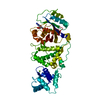
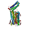
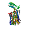
- PDB-4igb: Crystal structure of the N-terminal domain of the Streptococcus g... -
+ Open data

- Basic information
| Entry | Database: PDB / ID: 4igb | ||||||
|---|---|---|---|---|---|---|---|
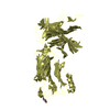
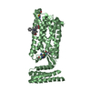
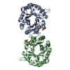
| Title | Crystal structure of the N-terminal domain of the Streptococcus gordonii adhesin Sgo0707 | ||||||
 Components Components | (LPXTG cell wall surface ...) x 4 | ||||||
 Keywords Keywords | CELL ADHESION / Beta-sandwich folds / Adhesin / Collagen-binding / oral keratinocytes / Cell wall anchored protein | ||||||
| Function / homology |  Function and homology information Function and homology informationSHIRT domain / Sgo0707, N-terminal domain / SHIRT domain / Sgo0707 N-terminal domain / Sgo0707-like, N2 domain / Sgo0707 N2 domain / Immunoglobulin-like - #740 / Fimbrial isopeptide formation D2 domain / LPXTG cell wall anchor motif / Gram-positive cocci surface proteins LPxTG motif profile. ...SHIRT domain / Sgo0707, N-terminal domain / SHIRT domain / Sgo0707 N-terminal domain / Sgo0707-like, N2 domain / Sgo0707 N2 domain / Immunoglobulin-like - #740 / Fimbrial isopeptide formation D2 domain / LPXTG cell wall anchor motif / Gram-positive cocci surface proteins LPxTG motif profile. / LPXTG cell wall anchor domain / Immunoglobulin-like / Sandwich / Mainly Beta Similarity search - Domain/homology | ||||||
| Biological species |  Streptococcus gordonii (bacteria) Streptococcus gordonii (bacteria) | ||||||
| Method |  X-RAY DIFFRACTION / SYNCHROTRON / SAD / Resolution: 2.09 Å X-RAY DIFFRACTION / SYNCHROTRON / SAD / Resolution: 2.09 Å | ||||||
 Authors Authors | Nylander, A. / Svensater, G. / Senadheera, D.B. / Cvitkovitch, D.G. / Davies, J.R. / Persson, K. | ||||||
 Citation Citation | Journal: Plos One / Year: 2013 Title: Structural and Functional Analysis of the N-terminal Domain of the Streptococcus gordonii Adhesin Sgo0707 Authors: Nylander, A. / Svensater, G. / Senadheera, D.B. / Cvitkovitch, D.G. / Davies, J.R. / Persson, K. | ||||||
| History |
|
- Structure visualization
| Structure viewer | Molecule: MolmilJmol/JSmol |
|---|
- Downloads & links
-Download
| PDBx/mmCIF format | 4igb.cif.gz | 719.5 KB | Display | PDBx/mmCIF format |
|---|---|---|---|---|
| PDB format | pdb4igb.ent.gz | 588 KB | Display | PDB format |
| PDBx/mmJSON format | 4igb.json.gz | Tree view | PDBx/mmJSON format | |
| Others |  Other downloads Other downloads |
-Validation report
| Arichive directory | https://data.pdbj.org/pub/pdb/validation_reports/ig/4igbftp://data.pdbj.org/pub/pdb/validation_reports/ig/4igb | HTTPS FTP |
|---|
-Related structure data
| Similar structure data |
|---|


-Links
 PDBj
PDBj- Assembly
| Deposited unit | 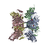
| ||||||||||||||||||||||||||||||
|---|---|---|---|---|---|---|---|---|---|---|---|---|---|---|---|---|---|---|---|---|---|---|---|---|---|---|---|---|---|---|---|
| 1 | 
| ||||||||||||||||||||||||||||||
| 2 | 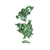
| ||||||||||||||||||||||||||||||
| 3 | 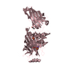
| ||||||||||||||||||||||||||||||
| 4 | 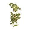
| ||||||||||||||||||||||||||||||
| Unit cell |
| ||||||||||||||||||||||||||||||
| Components on special symmetry positions |
| ||||||||||||||||||||||||||||||
| Noncrystallographic symmetry (NCS) | NCS domain:
NCS domain segments:
|
-Components
-LPXTG cell wall surface ... , 4 types, 4 molecules ABCD
| #1: Protein | Mass: 48054.316 Da / Num. of mol.: 1 / Fragment: Sgo0707-N, UNP residues 36-458 Source method: isolated from a genetically manipulated source Source: (gene. exp.) Streptococcus gordonii (bacteria) / Strain: Challis / ATCC 35105 / CH1 / DL1 / V288 / Gene: SGO_0707 / Plasmid: PetM11 / Production host: |
|---|---|
| #2: Protein | Mass: 48596.852 Da / Num. of mol.: 1 Source method: isolated from a genetically manipulated source Source: (gene. exp.) Streptococcus gordonii (bacteria) / Strain: Challis / ATCC 35105 / CH1 / DL1 / V288 / Gene: SGO_0707 / Plasmid: PetM11 / Production host: |
| #3: Protein | Mass: 48710.012 Da / Num. of mol.: 1 Source method: isolated from a genetically manipulated source Source: (gene. exp.) Streptococcus gordonii (bacteria) / Strain: Challis / ATCC 35105 / CH1 / DL1 / V288 / Gene: SGO_0707 / Plasmid: PetM11 / Production host: |
| #4: Protein | Mass: 47243.355 Da / Num. of mol.: 1 Source method: isolated from a genetically manipulated source Source: (gene. exp.) Streptococcus gordonii (bacteria) / Strain: Challis / ATCC 35105 / CH1 / DL1 / V288 / Gene: SGO_0707 / Plasmid: PetM11 / Production host: |
-Non-polymers , 5 types, 1300 molecules 


| #5: Chemical | ChemComp-SO4 /  Mass: 96.063 Da / Num. of mol.: 8 / Source method: obtained synthetically / Formula: SO4 Mass: 96.063 Da / Num. of mol.: 8 / Source method: obtained synthetically / Formula: SO4#6: Chemical | ChemComp-GOL /  Mass: 92.094 Da / Num. of mol.: 4 / Source method: obtained synthetically / Formula: C3H8O3 Mass: 92.094 Da / Num. of mol.: 4 / Source method: obtained synthetically / Formula: C3H8O3#7: Chemical |  Mass: 59.044 Da / Num. of mol.: 2 / Source method: obtained synthetically / Formula: C2H3O2 Mass: 59.044 Da / Num. of mol.: 2 / Source method: obtained synthetically / Formula: C2H3O2#8: Chemical | ChemComp-NA /  Mass: 22.990 Da / Num. of mol.: 4 / Source method: obtained synthetically / Formula: Na Mass: 22.990 Da / Num. of mol.: 4 / Source method: obtained synthetically / Formula: Na#9: Water | ChemComp-HOH / | Mass: 18.015 Da / Num. of mol.: 1282 / Source method: isolated from a natural source / Formula: H2O |
|---|
-Experimental details
-Experiment
| Experiment | Method: X-RAY DIFFRACTION / Number of used crystals: 2 |
|---|
- Sample preparation
| Crystal |
| |||||||||
|---|---|---|---|---|---|---|---|---|---|---|
| Crystal grow | Temperature: 291.15 K Details: 20%(w/v) PEG4000, 0.1M sodium acetate, 0.2M ammonium sulphate, pH 5.0, VAPOR DIFFUSION, SITTING DROP, temperature 291.15K PH range: 5.0; 5.0 |
-Data collection
| Diffraction |
| |||||||||||||||
|---|---|---|---|---|---|---|---|---|---|---|---|---|---|---|---|---|
| Diffraction source |
| |||||||||||||||
| Detector |
| |||||||||||||||
| Radiation |
| |||||||||||||||
| Radiation wavelength | Wavelength: 0.9793 Å / Relative weight: 1 | |||||||||||||||
| Reflection | Resolution: 2.09→48.45 Å / Num. obs: 114889 / % possible obs: 98.4 % / Observed criterion σ(I): -3 | |||||||||||||||
| Reflection shell | Resolution: 2.09→2.2 Å / % possible all: 98.4 |

- Processing
| Software |
| ||||||||||||||||||||||||||||||||||||||||||||||||||||||||||||||||||||||||||||||||||||||||||||||||||||||||||||||||||||||||||||||||||||||||||||||||||||||||||||||||||||||||||||||||||||||||||
|---|---|---|---|---|---|---|---|---|---|---|---|---|---|---|---|---|---|---|---|---|---|---|---|---|---|---|---|---|---|---|---|---|---|---|---|---|---|---|---|---|---|---|---|---|---|---|---|---|---|---|---|---|---|---|---|---|---|---|---|---|---|---|---|---|---|---|---|---|---|---|---|---|---|---|---|---|---|---|---|---|---|---|---|---|---|---|---|---|---|---|---|---|---|---|---|---|---|---|---|---|---|---|---|---|---|---|---|---|---|---|---|---|---|---|---|---|---|---|---|---|---|---|---|---|---|---|---|---|---|---|---|---|---|---|---|---|---|---|---|---|---|---|---|---|---|---|---|---|---|---|---|---|---|---|---|---|---|---|---|---|---|---|---|---|---|---|---|---|---|---|---|---|---|---|---|---|---|---|---|---|---|---|---|---|---|---|---|
| Refinement | Method to determine structure: SAD / Resolution: 2.09→41.07 Å / SU ML: 0.33 / σ(F): 0 / Phase error: 21.88 / Stereochemistry target values: ML
| ||||||||||||||||||||||||||||||||||||||||||||||||||||||||||||||||||||||||||||||||||||||||||||||||||||||||||||||||||||||||||||||||||||||||||||||||||||||||||||||||||||||||||||||||||||||||||
| Solvent computation | Shrinkage radii: 0.83 Å / VDW probe radii: 1.1 Å / Solvent model: FLAT BULK SOLVENT MODEL / Bsol: 42.48 Å2 / ksol: 0.37 e/Å3 | ||||||||||||||||||||||||||||||||||||||||||||||||||||||||||||||||||||||||||||||||||||||||||||||||||||||||||||||||||||||||||||||||||||||||||||||||||||||||||||||||||||||||||||||||||||||||||
| Displacement parameters |
| ||||||||||||||||||||||||||||||||||||||||||||||||||||||||||||||||||||||||||||||||||||||||||||||||||||||||||||||||||||||||||||||||||||||||||||||||||||||||||||||||||||||||||||||||||||||||||
| Refinement step | Cycle: LAST / Resolution: 2.09→41.07 Å
| ||||||||||||||||||||||||||||||||||||||||||||||||||||||||||||||||||||||||||||||||||||||||||||||||||||||||||||||||||||||||||||||||||||||||||||||||||||||||||||||||||||||||||||||||||||||||||
| Refine LS restraints |
| ||||||||||||||||||||||||||||||||||||||||||||||||||||||||||||||||||||||||||||||||||||||||||||||||||||||||||||||||||||||||||||||||||||||||||||||||||||||||||||||||||||||||||||||||||||||||||
| Refine LS restraints NCS |
| ||||||||||||||||||||||||||||||||||||||||||||||||||||||||||||||||||||||||||||||||||||||||||||||||||||||||||||||||||||||||||||||||||||||||||||||||||||||||||||||||||||||||||||||||||||||||||
| LS refinement shell | Refine-ID: X-RAY DIFFRACTION / Total num. of bins used: 30
| ||||||||||||||||||||||||||||||||||||||||||||||||||||||||||||||||||||||||||||||||||||||||||||||||||||||||||||||||||||||||||||||||||||||||||||||||||||||||||||||||||||||||||||||||||||||||||
| Refinement TLS params. | Method: refined / Origin x: 38.8013 Å / Origin y: -1.643 Å / Origin z: 41.6425 Å
| ||||||||||||||||||||||||||||||||||||||||||||||||||||||||||||||||||||||||||||||||||||||||||||||||||||||||||||||||||||||||||||||||||||||||||||||||||||||||||||||||||||||||||||||||||||||||||
| Refinement TLS group | Selection details: all |
