 Movie
Movie Controller

[English] 日本語
 Yorodumi
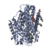
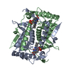
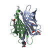
- PDB-5ko8: Crystal structure of haliscomenobacter hydrossis iodotyrosine dei... -
+ Open data

- Basic information
| Entry | Database: PDB / ID: 5ko8 | ||||||
|---|---|---|---|---|---|---|---|
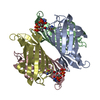
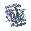
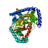
| Title | Crystal structure of haliscomenobacter hydrossis iodotyrosine deiodinase (IYD) bound to FMN and mono-iodotyrosine (I-Tyr) | ||||||
 Components Components | Nitroreductase | ||||||
 Keywords Keywords | OXIDOREDUCTASE / Haliscomenobacter hydrossis / iodotyrosine deiodinase / flavin mononucleotide | ||||||
| Function / homology |  Function and homology information Function and homology informationiodotyrosine deiodinase / iodotyrosine deiodinase activity / FMN binding Similarity search - Function | ||||||
| Biological species |  Haliscomenobacter hydrossis (bacteria) Haliscomenobacter hydrossis (bacteria) | ||||||
| Method |  X-RAY DIFFRACTION / SYNCHROTRON / MOLECULAR REPLACEMENT / Resolution: 2.15 Å X-RAY DIFFRACTION / SYNCHROTRON / MOLECULAR REPLACEMENT / Resolution: 2.15 Å | ||||||
 Authors Authors | Ingavat, N. / Kavran, J.M. / Sun, Z. / Rokita, S.E. | ||||||
 Citation Citation | Journal: Biochemistry / Year: 2017 Title: Active Site Binding Is Not Sufficient for Reductive Deiodination by Iodotyrosine Deiodinase. Authors: Ingavat, N. / Kavran, J.M. / Sun, Z. / Rokita, S.E. | ||||||
| History |
|
- Structure visualization
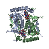
| Structure viewer | Molecule: MolmilJmol/JSmol |
|---|
- Downloads & links
-Download
| PDBx/mmCIF format | 5ko8.cif.gz | 181.7 KB | Display | PDBx/mmCIF format |
|---|---|---|---|---|
| PDB format | pdb5ko8.ent.gz | 147.3 KB | Display | PDB format |
| PDBx/mmJSON format | 5ko8.json.gz | Tree view | PDBx/mmJSON format | |
| Others |  Other downloads Other downloads |
-Validation report
| Summary document | 5ko8_validation.pdf.gz | 2.4 MB | Display | wwPDB validaton report |
|---|---|---|---|---|
| Full document | 5ko8_full_validation.pdf.gz | 2.4 MB | Display | |
| Data in XML | 5ko8_validation.xml.gz | 19.2 KB | Display | |
| Data in CIF | 5ko8_validation.cif.gz | 26 KB | Display | |
| Arichive directory | https://data.pdbj.org/pub/pdb/validation_reports/ko/5ko8ftp://data.pdbj.org/pub/pdb/validation_reports/ko/5ko8 | HTTPS FTP |
-Related structure data
| Related structure data | 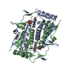 5ko7C 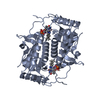 5krdC 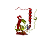 3gb5S C: citing same article ( S: Starting model for refinement |
|---|---|
| Similar structure data |


-Links
 PDBj
PDBj- Assembly
| Deposited unit | 
| ||||||||
|---|---|---|---|---|---|---|---|---|---|
| 1 | 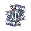
| ||||||||
| 2 | 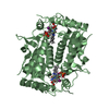
| ||||||||
| Unit cell |
|
-Components
| #1: Protein | Mass: 26165.963 Da / Num. of mol.: 2 Source method: isolated from a genetically manipulated source Source: (gene. exp.) Haliscomenobacter hydrossis (strain ATCC 27775 / DSM 1100 / LMG 10767 / O) (bacteria)Strain: ATCC 27775 / DSM 1100 / LMG 10767 / O / Gene: Halhy_2296 / Production host: #2: Chemical |   Mass: 456.344 Da / Num. of mol.: 2 / Source method: obtained synthetically / Formula: C17H21N4O9P Mass: 456.344 Da / Num. of mol.: 2 / Source method: obtained synthetically / Formula: C17H21N4O9P#3: Chemical | 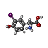  Type: L-peptide linking / Mass: 307.085 Da / Num. of mol.: 2 / Source method: obtained synthetically / Formula: C9H10INO3 Type: L-peptide linking / Mass: 307.085 Da / Num. of mol.: 2 / Source method: obtained synthetically / Formula: C9H10INO3#4: Water | ChemComp-HOH / |  Mass: 18.015 Da / Num. of mol.: 128 / Source method: isolated from a natural source / Formula: H2O Mass: 18.015 Da / Num. of mol.: 128 / Source method: isolated from a natural source / Formula: H2O |
|---|
-Experimental details
-Experiment
| Experiment | Method: X-RAY DIFFRACTION / Number of used crystals: 1 |
|---|
- Sample preparation
| Crystal | Density Matthews: 2.81 Å3/Da / Density % sol: 56.19 % |
|---|---|
| Crystal grow | Temperature: 298 K / Method: vapor diffusion, hanging drop / pH: 7.5 Details: 100 mM HEPES pH 7.5, 150 mM MgCl2, 25% (w/v) PEG 3350, 2 mM I-Tyr and 15% glycerol |
-Data collection
| Diffraction | Mean temperature: 100 K |
|---|---|
| Diffraction source | Source: SYNCHROTRON / Site: SSRL  / Beamline: BL12-2 / Wavelength: 0.9795 Å / Beamline: BL12-2 / Wavelength: 0.9795 Å |
| Detector | Type: DECTRIS PILATUS 6M / Detector: PIXEL / Date: Apr 22, 2015 |
| Radiation | Protocol: SINGLE WAVELENGTH / Monochromatic (M) / Laue (L): M / Scattering type: x-ray |
| Radiation wavelength | Wavelength: 0.9795 Å / Relative weight: 1 |
| Reflection | Resolution: 2.15→38.18 Å / Num. obs: 33077 / % possible obs: 99.9 % / Redundancy: 19.6 % / Rmerge(I) obs: 0.145 / Net I/σ(I): 16.8 |
- Processing
| Software |
| |||||||||||||||||||||||||||||||||||||||||||||||||||||||||||||||||||||||||||||||||||||||||||
|---|---|---|---|---|---|---|---|---|---|---|---|---|---|---|---|---|---|---|---|---|---|---|---|---|---|---|---|---|---|---|---|---|---|---|---|---|---|---|---|---|---|---|---|---|---|---|---|---|---|---|---|---|---|---|---|---|---|---|---|---|---|---|---|---|---|---|---|---|---|---|---|---|---|---|---|---|---|---|---|---|---|---|---|---|---|---|---|---|---|---|---|---|
| Refinement | Method to determine structure: MOLECULAR REPLACEMENT Starting model: 3GB5 Resolution: 2.15→38.18 Å / SU ML: 0.31 / Cross valid method: FREE R-VALUE / σ(F): 1.33 / Phase error: 25.49 / Stereochemistry target values: ML
| |||||||||||||||||||||||||||||||||||||||||||||||||||||||||||||||||||||||||||||||||||||||||||
| Solvent computation | Shrinkage radii: 0.9 Å / VDW probe radii: 1.11 Å / Solvent model: FLAT BULK SOLVENT MODEL | |||||||||||||||||||||||||||||||||||||||||||||||||||||||||||||||||||||||||||||||||||||||||||
| Refinement step | Cycle: LAST / Resolution: 2.15→38.18 Å
| |||||||||||||||||||||||||||||||||||||||||||||||||||||||||||||||||||||||||||||||||||||||||||
| Refine LS restraints |
| |||||||||||||||||||||||||||||||||||||||||||||||||||||||||||||||||||||||||||||||||||||||||||
| LS refinement shell |
|
