 Movie
Movie Controller


+ Open data

- Basic information
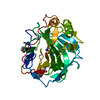
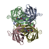
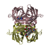
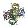
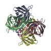
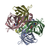
| Entry | Database: PDB / ID: 1luq | ||||||
|---|---|---|---|---|---|---|---|
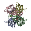
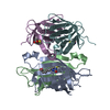
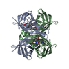
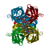
| Title | Full Matrix Error Analysis of Streptavidin | ||||||
 Components Components | Streptavidin | ||||||
 Keywords Keywords | BINDING PROTEIN | ||||||
| Function / homology |  Function and homology information Function and homology information | ||||||
| Biological species |  Streptomyces avidinii (bacteria) Streptomyces avidinii (bacteria) | ||||||
| Method |  X-RAY DIFFRACTION / SYNCHROTRON / MOLECULAR REPLACEMENT / Resolution: 0.96 Å X-RAY DIFFRACTION / SYNCHROTRON / MOLECULAR REPLACEMENT / Resolution: 0.96 Å | ||||||
 Authors Authors | Merritt, E.A. / Le Trong, I. | ||||||
 Citation Citation | Journal: To be Published Title: Full Matrix Error Analysis of Streptavidin Authors: Merritt, E.A. / Le Trong, I. | ||||||
| History |
|
- Structure visualization
| Structure viewer | Molecule: MolmilJmol/JSmol |
|---|
- Downloads & links
-Download
| PDBx/mmCIF format | 1luq.cif.gz | 151.5 KB | Display | PDBx/mmCIF format |
|---|---|---|---|---|
| PDB format | pdb1luq.ent.gz | 145.4 KB | Display | PDB format |
| PDBx/mmJSON format | 1luq.json.gz | Tree view | PDBx/mmJSON format | |
| Others |  Other downloads Other downloads |
-Validation report
| Arichive directory | https://data.pdbj.org/pub/pdb/validation_reports/lu/1luqftp://data.pdbj.org/pub/pdb/validation_reports/lu/1luq | HTTPS FTP |
|---|
-Related structure data
| Related structure data |  1df8S S: Starting model for refinement |
|---|---|
| Similar structure data |
-Links
 PDBj
PDBj- Assembly
| Deposited unit | 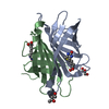
| |||||||||||||||
|---|---|---|---|---|---|---|---|---|---|---|---|---|---|---|---|---|
| 1 | 
| |||||||||||||||
| Unit cell |
| |||||||||||||||
| Components on special symmetry positions |
|


-Components
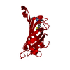
| #1: Protein | Mass: 13265.336 Da / Num. of mol.: 2 / Mutation: S1045A Source method: isolated from a genetically manipulated source Source: (gene. exp.) Streptomyces avidinii (bacteria) / Description: T7 expression system / Production host: #2: Chemical |   Mass: 244.311 Da / Num. of mol.: 2 / Source method: obtained synthetically / Formula: C10H16N2O3S Mass: 244.311 Da / Num. of mol.: 2 / Source method: obtained synthetically / Formula: C10H16N2O3S#3: Chemical | ChemComp-GOL / |   Mass: 92.094 Da / Num. of mol.: 1 / Source method: obtained synthetically / Formula: C3H8O3 Mass: 92.094 Da / Num. of mol.: 1 / Source method: obtained synthetically / Formula: C3H8O3#4: Chemical | ChemComp-MPD / ( |   Mass: 118.174 Da / Num. of mol.: 1 / Source method: obtained synthetically / Formula: C6H14O2 / Comment: precipitant*YM Mass: 118.174 Da / Num. of mol.: 1 / Source method: obtained synthetically / Formula: C6H14O2 / Comment: precipitant*YM#5: Water | ChemComp-HOH / |  Mass: 18.015 Da / Num. of mol.: 302 / Source method: isolated from a natural source / Formula: H2O Mass: 18.015 Da / Num. of mol.: 302 / Source method: isolated from a natural source / Formula: H2O |
|---|
-Experimental details
-Experiment
| Experiment | Method: X-RAY DIFFRACTION / Number of used crystals: 1 |
|---|
- Sample preparation
| Crystal | Density Matthews: 1.89 Å3/Da / Density meas: 34.8 Mg/m3 / Density % sol: 41.78 % |
|---|---|
| Crystal grow | Temperature: 293 K / Method: vapor diffusion, sitting drop / pH: 4.5 Details: ammonium sulfate, sodium acetate, biotin, pH 4.5, VAPOR DIFFUSION, SITTING DROP, temperature 293K |
-Data collection
| Diffraction | Mean temperature: 100 K |
|---|---|
| Diffraction source | Source: SYNCHROTRON / Site: SSRL  / Beamline: BL9-1 / Wavelength: 0.98 / Beamline: BL9-1 / Wavelength: 0.98 |
| Detector | Type: MARRESEARCH / Detector: IMAGE PLATE / Date: Jan 1, 1999 |
| Radiation | Protocol: SINGLE WAVELENGTH / Monochromatic (M) / Laue (L): M / Scattering type: x-ray |
| Radiation wavelength | Wavelength: 0.98 Å / Relative weight: 1 |
| Reflection | Resolution: 0.96→14 Å / Num. all: 135985 / Num. obs: 135985 / % possible obs: 96.4 % / Observed criterion σ(F): 0 / Observed criterion σ(I): 0 / Redundancy: 8.9 % / Rmerge(I) obs: 0.065 |
| Reflection shell | Resolution: 0.96→0.97 Å / Rmerge(I) obs: 0.602 / % possible all: 45 |
- Processing
| Software |
| |||||||||||||||||||||||||||||||||
|---|---|---|---|---|---|---|---|---|---|---|---|---|---|---|---|---|---|---|---|---|---|---|---|---|---|---|---|---|---|---|---|---|---|---|
| Refinement | Method to determine structure: MOLECULAR REPLACEMENT Starting model: 1DF8 Resolution: 0.96→14 Å / Num. parameters: 19886 / Num. restraintsaints: 25548 / Cross valid method: FREE R / σ(F): 0 / Stereochemistry target values: Engh & Huber Details: ANISOTROPIC REFINEMENT REDUCED FREE R (NO CUTOFF) BY 4.0%. Entry 1LUQ contains the crystallographic model used for error analysis and model comparison as reported in Merritt et al. The final ...Details: ANISOTROPIC REFINEMENT REDUCED FREE R (NO CUTOFF) BY 4.0%. Entry 1LUQ contains the crystallographic model used for error analysis and model comparison as reported in Merritt et al. The final crystallographic model for this structure will be deposited separately under a different accession code.
| |||||||||||||||||||||||||||||||||
| Solvent computation | Solvent model: MOEWS & KRETSINGER, J.MOL.BIOL.91(1973)201-228 | |||||||||||||||||||||||||||||||||
| Refine analyze | Num. disordered residues: 14 / Occupancy sum hydrogen: 1667.5 / Occupancy sum non hydrogen: 2128 | |||||||||||||||||||||||||||||||||
| Refinement step | Cycle: LAST / Resolution: 0.96→14 Å
| |||||||||||||||||||||||||||||||||
| Refine LS restraints |
|
