 Movie
Movie Controller

[English] 日本語
 Yorodumi
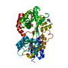
- PDB-5khl: Crystal Structure of periplasmic Heme binding protein HutB of Vib... -
+ Open data

- Basic information
| Entry | Database: PDB / ID: 5khl | ||||||
|---|---|---|---|---|---|---|---|
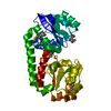
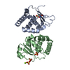
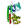
| Title | Crystal Structure of periplasmic Heme binding protein HutB of Vibrio cholerae | ||||||
 Components Components | Hemin ABC transporter, periplasmic hemin-binding protein HutB | ||||||
 Keywords Keywords | TRANSPORT PROTEIN / Hemin / ABC trasporter / periplasmic protein / Vibrio cholerae | ||||||
| Function / homology | : / ABC transporter periplasmic binding domain / Periplasmic binding protein / Iron siderophore/cobalamin periplasmic-binding domain profile. / Nitrogenase molybdenum iron protein domain / Rossmann fold / 3-Layer(aba) Sandwich / Alpha Beta / Hemin ABC transporter, periplasmic hemin-binding protein HutB Function and homology information Function and homology information | ||||||
| Biological species |   Vibrio cholerae (bacteria) Vibrio cholerae (bacteria) | ||||||
| Method |  X-RAY DIFFRACTION / SYNCHROTRON / MOLECULAR REPLACEMENT / Resolution: 2.397 Å X-RAY DIFFRACTION / SYNCHROTRON / MOLECULAR REPLACEMENT / Resolution: 2.397 Å | ||||||
 Authors Authors | Agarwal, S. / Dey, S. / Ghosh, B. / Dasgupta, J. | ||||||
| Funding support |  India, 1items India, 1items
| ||||||
 Citation Citation | Journal: To be published Title: Crystal Structure of periplasmic Heme binding protein HutB of Vibrio cholerae Authors: Agarwal, S. / Biswas, M. / Dasgupta, J. #1: Journal: Acta Crystallogr F Struct Biol Commun / Year: 2015 Title: Purification, crystallization and preliminary X-ray analysis of the periplasmic haem-binding protein HutB from Vibrio cholerae. Authors: Agarwal, S. / Biswas, M. / Dasgupta, J. | ||||||
| History |
|
- Structure visualization
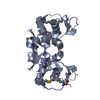
| Structure viewer | Molecule: MolmilJmol/JSmol |
|---|
- Downloads & links
-Download
| PDBx/mmCIF format | 5khl.cif.gz | 111.7 KB | Display | PDBx/mmCIF format |
|---|---|---|---|---|
| PDB format | pdb5khl.ent.gz | 85.8 KB | Display | PDB format |
| PDBx/mmJSON format | 5khl.json.gz | Tree view | PDBx/mmJSON format | |
| Others |  Other downloads Other downloads |
-Validation report
| Arichive directory | https://data.pdbj.org/pub/pdb/validation_reports/kh/5khlftp://data.pdbj.org/pub/pdb/validation_reports/kh/5khl | HTTPS FTP |
|---|
-Related structure data
| Related structure data | 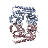 2rg7S S: Starting model for refinement |
|---|---|
| Similar structure data |


-Links
 PDBj
PDBj- Assembly
| Deposited unit | 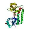
| ||||||||
|---|---|---|---|---|---|---|---|---|---|
| 1 |
| ||||||||
| Unit cell |
|
-Components
| #1: Protein | Mass: 28860.943 Da / Num. of mol.: 1 / Fragment: UNP RESIDUES 24-277 Source method: isolated from a genetically manipulated source Source: (gene. exp.) Vibrio cholerae (bacteria) / Strain: O395 / Gene: hutB, VC0395_0324 / Production host: |
|---|---|
| #2: Chemical | ChemComp-SO4 /   Mass: 96.063 Da / Num. of mol.: 1 / Source method: obtained synthetically / Formula: SO4 Mass: 96.063 Da / Num. of mol.: 1 / Source method: obtained synthetically / Formula: SO4 |
| #3: Water | ChemComp-HOH /  Mass: 18.015 Da / Num. of mol.: 48 / Source method: isolated from a natural source / Formula: H2O Mass: 18.015 Da / Num. of mol.: 48 / Source method: isolated from a natural source / Formula: H2O |
-Experimental details
-Experiment
| Experiment | Method: X-RAY DIFFRACTION / Number of used crystals: 1 |
|---|
- Sample preparation
| Crystal | Density Matthews: 2.33 Å3/Da / Density % sol: 47.11 % |
|---|---|
| Crystal grow | Temperature: 293 K / Method: vapor diffusion, hanging drop / pH: 7 Details: 0.8 M ammonium sulfate, 0.1 M HEPES (pH 7.0) against a reservoir solution 1.6 M ammonium sulfate |
-Data collection
| Diffraction | Mean temperature: 100 K |
|---|---|
| Diffraction source | Source: SYNCHROTRON / Site: RRCAT INDUS-2 / Beamline: PX-BL21 / Wavelength: 0.98 Å |
| Detector | Type: RAYONIX MX-225 / Detector: CCD / Date: Aug 24, 2014 |
| Radiation | Protocol: SINGLE WAVELENGTH / Monochromatic (M) / Laue (L): M / Scattering type: x-ray |
| Radiation wavelength | Wavelength: 0.98 Å / Relative weight: 1 |
| Reflection | Resolution: 2.397→37 Å / Num. obs: 11192 / % possible obs: 99.2 % / Redundancy: 9.7 % / Net I/σ(I): 29.9 |
| Reflection shell | Resolution: 2.4→2.49 Å |
- Processing
| Software |
| |||||||||||||||||||||||||||||||||||||||||||||||||||||||||||||||||||||||||||
|---|---|---|---|---|---|---|---|---|---|---|---|---|---|---|---|---|---|---|---|---|---|---|---|---|---|---|---|---|---|---|---|---|---|---|---|---|---|---|---|---|---|---|---|---|---|---|---|---|---|---|---|---|---|---|---|---|---|---|---|---|---|---|---|---|---|---|---|---|---|---|---|---|---|---|---|---|
| Refinement | Method to determine structure: MOLECULAR REPLACEMENT Starting model: 2RG7 Resolution: 2.397→28.53 Å / SU ML: 0.37 / Cross valid method: FREE R-VALUE / σ(F): 1.34 / Phase error: 28.12 / Stereochemistry target values: ML
| |||||||||||||||||||||||||||||||||||||||||||||||||||||||||||||||||||||||||||
| Solvent computation | Shrinkage radii: 0.9 Å / VDW probe radii: 1.11 Å / Solvent model: FLAT BULK SOLVENT MODEL | |||||||||||||||||||||||||||||||||||||||||||||||||||||||||||||||||||||||||||
| Refinement step | Cycle: LAST / Resolution: 2.397→28.53 Å
| |||||||||||||||||||||||||||||||||||||||||||||||||||||||||||||||||||||||||||
| Refine LS restraints |
| |||||||||||||||||||||||||||||||||||||||||||||||||||||||||||||||||||||||||||
| LS refinement shell |
| |||||||||||||||||||||||||||||||||||||||||||||||||||||||||||||||||||||||||||
| Refinement TLS params. | Method: refined / Refine-ID: X-RAY DIFFRACTION
| |||||||||||||||||||||||||||||||||||||||||||||||||||||||||||||||||||||||||||
| Refinement TLS group |
|
