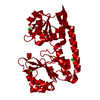
- PDB-2wi8: Crystal structure of the triscatecholate siderophore binding prot... -
+
Open data
ID or keywords:
Loading...
-
Basic information
Entry
Database: PDB / ID: 2wi8
Title
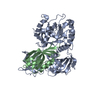
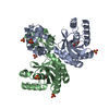
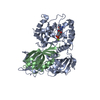
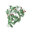
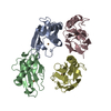
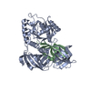
Crystal structure of the triscatecholate siderophore binding protein FeuA from Bacillus subtilis
Components
IRON-UPTAKE SYSTEM-BINDING PROTEIN
Keywords
TRANSPORT PROTEIN / BACILLIBACTIN AND ENTEROBACTIN BINDING / TRISCATECHOLATE BINDING PROTEIN / IRON TRANSPORT / HIGH AFFINITY IRON IMPORT / IRON / MEMBRANE / PALMITATE / TRANSPORT / ABC-TYPE TRANSPORTER BINDING PROTEIN / SIDEROPHORE BINDING PROTEIN / LIPOPROTEIN / CELL MEMBRANE / ION TRANSPORT
Function / homology
Function and homology information
iron coordination entity transport / outer membrane-bounded periplasmic space / membrane raft / plasma membrane / cytoplasm Similarity search - Function
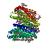
: / ABC transporter periplasmic binding domain / Periplasmic binding protein / Iron siderophore/cobalamin periplasmic-binding domain profile. / Nitrogenase molybdenum iron protein domain / Prokaryotic membrane lipoprotein lipid attachment site profile. / Rossmann fold / 3-Layer(aba) Sandwich / Alpha Beta Similarity search - Domain/homology
Mass: 18.015 Da / Num. of mol.: 239 / Source method: isolated from a natural source / Formula: H2O
Sequence details
I180T S237I ARE SEQUENCE VARIANTS
-
Experimental details
-
Experiment
Experiment
Method: X-RAY DIFFRACTION / Number of used crystals: 1
-
Sample preparation
Crystal
Density Matthews: 1.92 Å3/Da / Density % sol: 35.88 % / Description: NONE
Crystal grow
pH: 8 Details: PROTEIN WAS CRYSTALLIZED FROM 30% (V/V) JEFFAMINE ED-2001 PH 7.0, 100 MM HEPES, PH 8.0; THEN SOAKED IN MOTHER LIQUOR CONTAINING 30% (V/V) GLYCEROL FOR CRYOPROTECTION
 Movie
Movie Controller

 Yorodumi
 Open data
 Basic information
 Components
 Keywords
 Function and homology information
 X-RAY DIFFRACTION /
  Authors
 Citation
 Structure visualization
 Downloads & links
 Other downloads


 PDBj
PDBj


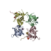
 Assembly

 Mass: 35.453 Da / Num. of mol.: 3 / Source method: obtained synthetically / Formula: Cl
 Mass: 18.015 Da / Num. of mol.: 239 / Source method: isolated from a natural source / Formula: H2O
 Sample preparation
 / Beamline: ID29 / Wavelength: 0.97623
  Processing