 Movie
Movie Controller

[English] 日本語
 Yorodumi
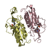
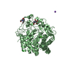
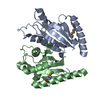
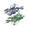
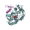
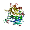
- PDB-5kap: Trypanosome brucei Hypoxanthine-guanine phosphoribosyltranferase ... -
+ Open data

- Basic information
| Entry | Database: PDB / ID: 5kap | ||||||
|---|---|---|---|---|---|---|---|
| Title | Trypanosome brucei Hypoxanthine-guanine phosphoribosyltranferase in complex with a 9-(4-(phosphonobutil)hypoxanthine | ||||||
 Components Components | Hypoxanthine-guanine phosphoribosyltransferase | ||||||
 Keywords Keywords | TRANSFERASE/TRANSFERASE INHIBITOR / inhibitor / complex / dimer / enzyme / TRANSFERASE / TRANSFERASE-TRANSFERASE INHIBITOR complex | ||||||
| Function / homology |  Function and homology information Function and homology informationhypoxanthine phosphoribosyltransferase / guanine phosphoribosyltransferase activity / guanine salvage / hypoxanthine metabolic process / hypoxanthine phosphoribosyltransferase activity / GMP salvage / IMP salvage / glycosome / nuclear lumen / ciliary plasm ...hypoxanthine phosphoribosyltransferase / guanine phosphoribosyltransferase activity / guanine salvage / hypoxanthine metabolic process / hypoxanthine phosphoribosyltransferase activity / GMP salvage / IMP salvage / glycosome / nuclear lumen / ciliary plasm / purine ribonucleoside salvage / nucleotide binding / magnesium ion binding / cytosol / cytoplasm Similarity search - Function | ||||||
| Biological species |  | ||||||
| Method |  X-RAY DIFFRACTION / SYNCHROTRON / MOLECULAR REPLACEMENT / Resolution: 2.95 Å X-RAY DIFFRACTION / SYNCHROTRON / MOLECULAR REPLACEMENT / Resolution: 2.95 Å | ||||||
 Authors Authors | Teran, D. / Guddat, L. | ||||||
 Citation Citation | Journal: Sci Rep / Year: 2016 Title: Crystal structures and inhibition of Trypanosoma brucei hypoxanthine-guanine phosphoribosyltransferase. Authors: Teran, D. / Hockova, D. / Cesnek, M. / Zikova, A. / Naesens, L. / Keough, D.T. / Guddat, L.W. | ||||||
| History |
|
- Structure visualization
| Structure viewer | Molecule: MolmilJmol/JSmol |
|---|
- Downloads & links
-Download
| PDBx/mmCIF format | 5kap.cif.gz | 152.9 KB | Display | PDBx/mmCIF format |
|---|---|---|---|---|
| PDB format | pdb5kap.ent.gz | 120.1 KB | Display | PDB format |
| PDBx/mmJSON format | 5kap.json.gz | Tree view | PDBx/mmJSON format | |
| Others |  Other downloads Other downloads |
-Validation report
| Arichive directory | https://data.pdbj.org/pub/pdb/validation_reports/ka/5kapftp://data.pdbj.org/pub/pdb/validation_reports/ka/5kap | HTTPS FTP |
|---|
-Related structure data
| Related structure data | 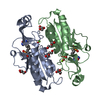 5jsqSC 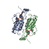 5jv5C 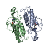 5k51C 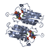 5kamC C: citing same article ( S: Starting model for refinement |
|---|---|
| Similar structure data |


-Links
 PDBj
PDBj
- Assembly
| Deposited unit | 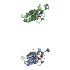
| ||||||||
|---|---|---|---|---|---|---|---|---|---|
| 1 | 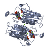
| ||||||||
| 2 | 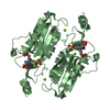
| ||||||||
| Unit cell |
| ||||||||
| Components on special symmetry positions |
|
-Components
| #1: Protein | Mass: 24221.775 Da / Num. of mol.: 2 Source method: isolated from a genetically manipulated source Source: (gene. exp.)  References: UniProt: Q07010, hypoxanthine phosphoribosyltransferase #2: Chemical | 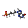  Mass: 272.198 Da / Num. of mol.: 2 / Source method: obtained synthetically / Formula: C9H13N4O4P Mass: 272.198 Da / Num. of mol.: 2 / Source method: obtained synthetically / Formula: C9H13N4O4P#3: Chemical |   Mass: 96.063 Da / Num. of mol.: 3 / Source method: obtained synthetically / Formula: SO4 Mass: 96.063 Da / Num. of mol.: 3 / Source method: obtained synthetically / Formula: SO4#4: Chemical | ChemComp-MG /   Mass: 24.305 Da / Num. of mol.: 6 / Source method: obtained synthetically / Formula: Mg Mass: 24.305 Da / Num. of mol.: 6 / Source method: obtained synthetically / Formula: Mg#5: Water | ChemComp-HOH / |  Mass: 18.015 Da / Num. of mol.: 12 / Source method: isolated from a natural source / Formula: H2O Mass: 18.015 Da / Num. of mol.: 12 / Source method: isolated from a natural source / Formula: H2O |
|---|
-Experimental details
-Experiment
| Experiment | Method: X-RAY DIFFRACTION / Number of used crystals: 1 |
|---|
- Sample preparation
| Crystal | Density Matthews: 2.4 Å3/Da / Density % sol: 48.74 % |
|---|---|
| Crystal grow | Temperature: 293.15 K / Method: vapor diffusion, hanging drop Details: 25% PEG 3350, 0.2 M lithium sulfate and 0.1 M Bis-Tris PH range: 5-5.5 |
-Data collection
| Diffraction | Mean temperature: 100 K |
|---|---|
| Diffraction source | Source: SYNCHROTRON / Site: Australian Synchrotron  / Beamline: MX1 / Wavelength: 0.95369 Å / Beamline: MX1 / Wavelength: 0.95369 Å |
| Detector | Type: ADSC QUANTUM 270 / Detector: CCD / Date: Jun 5, 2015 |
| Radiation | Protocol: SINGLE WAVELENGTH / Monochromatic (M) / Laue (L): M / Scattering type: x-ray |
| Radiation wavelength | Wavelength: 0.95369 Å / Relative weight: 1 |
| Reflection | Resolution: 2.81→47.39 Å / Num. obs: 11686 / % possible obs: 98.8 % / Redundancy: 7.3 % / Rmerge(I) obs: 0.105 / Net I/σ(I): 13 |
| Reflection shell | Resolution: 2.81→2.97 Å / Redundancy: 7.4 % / Rmerge(I) obs: 0.71 / Mean I/σ(I) obs: 2.6 / % possible all: 97.4 |
- Processing
| Software |
| ||||||||||||||||||||||||||||||||||||||||||||||||||||||||
|---|---|---|---|---|---|---|---|---|---|---|---|---|---|---|---|---|---|---|---|---|---|---|---|---|---|---|---|---|---|---|---|---|---|---|---|---|---|---|---|---|---|---|---|---|---|---|---|---|---|---|---|---|---|---|---|---|---|
| Refinement | Method to determine structure: MOLECULAR REPLACEMENT Starting model: 5JSQ Resolution: 2.95→45.106 Å / SU ML: 0.44 / Cross valid method: FREE R-VALUE / σ(F): 1.36 / Phase error: 35.56 / Stereochemistry target values: ML
| ||||||||||||||||||||||||||||||||||||||||||||||||||||||||
| Solvent computation | Shrinkage radii: 0.9 Å / VDW probe radii: 1.11 Å / Solvent model: FLAT BULK SOLVENT MODEL | ||||||||||||||||||||||||||||||||||||||||||||||||||||||||
| Refinement step | Cycle: LAST / Resolution: 2.95→45.106 Å
| ||||||||||||||||||||||||||||||||||||||||||||||||||||||||
| Refine LS restraints |
| ||||||||||||||||||||||||||||||||||||||||||||||||||||||||
| LS refinement shell |
| ||||||||||||||||||||||||||||||||||||||||||||||||||||||||
| Refinement TLS params. | Method: refined / Origin x: -36.4196 Å / Origin y: 32.7608 Å / Origin z: 23.6149 Å
| ||||||||||||||||||||||||||||||||||||||||||||||||||||||||
| Refinement TLS group | Selection details: all |
