 Movie
Movie Controller


+ Open data

- Basic information
| Entry | Database: PDB / ID: 5jod | |||||||||
|---|---|---|---|---|---|---|---|---|---|---|
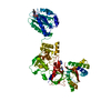
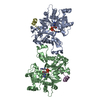
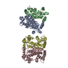
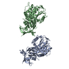
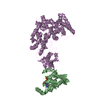
| Title | Structure of proplasmepsin IV from Plasmodium falciparum | |||||||||
 Components Components | Proplasmepsin IV | |||||||||
 Keywords Keywords | HYDROLASE / malaria | |||||||||
| Function / homology |  Function and homology information Function and homology informationvacuolar lumen / food vacuole / aspartic-type endopeptidase activity / proteolysis / membrane Similarity search - Function | |||||||||
| Biological species |  | |||||||||
| Method |  X-RAY DIFFRACTION / SYNCHROTRON / MOLECULAR REPLACEMENT / molecular replacement / Resolution: 1.528 Å X-RAY DIFFRACTION / SYNCHROTRON / MOLECULAR REPLACEMENT / molecular replacement / Resolution: 1.528 Å | |||||||||
 Authors Authors | Recacha, R. / Akopjana, I. / Tars, K. / Jaudzems, K. | |||||||||
 Citation Citation | Journal: Acta Crystallogr.,Sect.F / Year: 2016 Title: Crystal structure of Plasmodium falciparum proplasmepsin IV: the plasticity of proplasmepsins. Authors: Recacha, R. / Jaudzems, K. / Akopjana, I. / Jirgensons, A. / Tars, K. #1: Journal: Nat. Struct. Biol. / Year: 1999Title: Crystal structure of the novel aspartic proteinase zymogen proplasmepsin II from plasmodium falciparum. Authors: Bernstein, N.K. / Cherney, M.M. / Loetscher, H. / Ridley, R.G. / James, M.N. #2: Journal: J. Mol. Biol. / Year: 2003Title: Structural insights into the activation of P. vivax plasmepsin. Authors: Bernstein, N.K. / Cherney, M.M. / Yowell, C.A. / Dame, J.B. / James, M.N. | |||||||||
| History |
|
- Structure visualization
| Structure viewer | Molecule: MolmilJmol/JSmol |
|---|
- Downloads & links
-Download
| PDBx/mmCIF format | 5jod.cif.gz | 321.7 KB | Display | PDBx/mmCIF format |
|---|---|---|---|---|
| PDB format | pdb5jod.ent.gz | 261.4 KB | Display | PDB format |
| PDBx/mmJSON format | 5jod.json.gz | Tree view | PDBx/mmJSON format | |
| Others |  Other downloads Other downloads |
-Validation report
| Arichive directory | https://data.pdbj.org/pub/pdb/validation_reports/jo/5jodftp://data.pdbj.org/pub/pdb/validation_reports/jo/5jod | HTTPS FTP |
|---|
-Related structure data
| Related structure data |  5bwyC  1pfzS S: Starting model for refinement C: citing same article ( |
|---|---|
| Similar structure data |
-Links
 PDBj
PDBj- Assembly
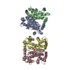
| Deposited unit | 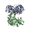
| ||||||||
|---|---|---|---|---|---|---|---|---|---|
| 1 | 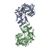
| ||||||||
| 2 | 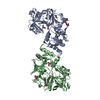
| ||||||||
| Unit cell |
|
-Components
| #1: Protein | Mass: 42372.699 Da / Num. of mol.: 2 / Fragment: UNP residues 75-449 Source method: isolated from a genetically manipulated source Source: (gene. exp.) Gene: PFBG_05102 / Production host:  #2: Chemical | ChemComp-GOL /   Mass: 92.094 Da / Num. of mol.: 9 / Source method: obtained synthetically / Formula: C3H8O3 Mass: 92.094 Da / Num. of mol.: 9 / Source method: obtained synthetically / Formula: C3H8O3#3: Water | ChemComp-HOH / |  Mass: 18.015 Da / Num. of mol.: 689 / Source method: isolated from a natural source / Formula: H2O Mass: 18.015 Da / Num. of mol.: 689 / Source method: isolated from a natural source / Formula: H2OHas protein modification | Y | |
|---|
-Experimental details
-Experiment
| Experiment | Method: X-RAY DIFFRACTION / Number of used crystals: 1 |
|---|
- Sample preparation
| Crystal | Density Matthews: 2.65 Å3/Da / Density % sol: 53.55 % |
|---|---|
| Crystal grow | Temperature: 293 K / Method: vapor diffusion, sitting drop / pH: 4.7 Details: 100 mM sodium citrate, 25% (w/v) PEG 3350 200 mM ammonium acetate |
-Data collection
| Diffraction | Mean temperature: 100 K |
|---|---|
| Diffraction source | Source: SYNCHROTRON / Site: MAX II  / Beamline: I911-3 / Wavelength: 0.97 Å / Beamline: I911-3 / Wavelength: 0.97 Å |
| Detector | Type: ADSC QUANTUM 210 / Detector: CCD / Date: May 24, 2014 |
| Radiation | Protocol: SINGLE WAVELENGTH / Monochromatic (M) / Laue (L): M / Scattering type: x-ray |
| Radiation wavelength | Wavelength: 0.97 Å / Relative weight: 1 |
| Reflection twin | Operator: -h,-k,h+l / Fraction: 0.04 |
| Reflection | Resolution: 1.53→48.41 Å / Num. obs: 129149 / % possible obs: 96.3 % / Redundancy: 2.7 % / CC1/2: 0.995 / Rmerge(I) obs: 0.065 / Net I/av σ(I): 7.5 / Net I/σ(I): 7.5 |
| Reflection shell | Resolution: 1.53→1.55 Å / Redundancy: 2.1 % / Rmerge(I) obs: 0.609 / Mean I/σ(I) obs: 1.2 / % possible all: 95.1 |
-Phasing
| Phasing | Method: molecular replacement |
|---|
- Processing
| Software |
| |||||||||||||||||||||||||||||||||||||||||||||||||||||||||||||||||||||||||||||||||||||||||||||||||||||||||||||||||||||||||||||||||||||||||||||||||||||||||||||||||||||||||||||||||||||||||||||||||||||||||||||||||||||||||
|---|---|---|---|---|---|---|---|---|---|---|---|---|---|---|---|---|---|---|---|---|---|---|---|---|---|---|---|---|---|---|---|---|---|---|---|---|---|---|---|---|---|---|---|---|---|---|---|---|---|---|---|---|---|---|---|---|---|---|---|---|---|---|---|---|---|---|---|---|---|---|---|---|---|---|---|---|---|---|---|---|---|---|---|---|---|---|---|---|---|---|---|---|---|---|---|---|---|---|---|---|---|---|---|---|---|---|---|---|---|---|---|---|---|---|---|---|---|---|---|---|---|---|---|---|---|---|---|---|---|---|---|---|---|---|---|---|---|---|---|---|---|---|---|---|---|---|---|---|---|---|---|---|---|---|---|---|---|---|---|---|---|---|---|---|---|---|---|---|---|---|---|---|---|---|---|---|---|---|---|---|---|---|---|---|---|---|---|---|---|---|---|---|---|---|---|---|---|---|---|---|---|---|---|---|---|---|---|---|---|---|---|---|---|---|---|---|---|---|
| Refinement | Method to determine structure: MOLECULAR REPLACEMENT Starting model: 1PFZ Resolution: 1.528→36.679 Å / SU ML: 0.2 / Cross valid method: FREE R-VALUE / σ(F): 1.34 / Phase error: 23.91 / Stereochemistry target values: ML
| |||||||||||||||||||||||||||||||||||||||||||||||||||||||||||||||||||||||||||||||||||||||||||||||||||||||||||||||||||||||||||||||||||||||||||||||||||||||||||||||||||||||||||||||||||||||||||||||||||||||||||||||||||||||||
| Solvent computation | Shrinkage radii: 0.9 Å / VDW probe radii: 1.11 Å / Solvent model: FLAT BULK SOLVENT MODEL | |||||||||||||||||||||||||||||||||||||||||||||||||||||||||||||||||||||||||||||||||||||||||||||||||||||||||||||||||||||||||||||||||||||||||||||||||||||||||||||||||||||||||||||||||||||||||||||||||||||||||||||||||||||||||
| Refinement step | Cycle: LAST / Resolution: 1.528→36.679 Å
| |||||||||||||||||||||||||||||||||||||||||||||||||||||||||||||||||||||||||||||||||||||||||||||||||||||||||||||||||||||||||||||||||||||||||||||||||||||||||||||||||||||||||||||||||||||||||||||||||||||||||||||||||||||||||
| Refine LS restraints |
| |||||||||||||||||||||||||||||||||||||||||||||||||||||||||||||||||||||||||||||||||||||||||||||||||||||||||||||||||||||||||||||||||||||||||||||||||||||||||||||||||||||||||||||||||||||||||||||||||||||||||||||||||||||||||
| LS refinement shell |
| |||||||||||||||||||||||||||||||||||||||||||||||||||||||||||||||||||||||||||||||||||||||||||||||||||||||||||||||||||||||||||||||||||||||||||||||||||||||||||||||||||||||||||||||||||||||||||||||||||||||||||||||||||||||||
| Refinement TLS params. | Method: refined / Refine-ID: X-RAY DIFFRACTION
| |||||||||||||||||||||||||||||||||||||||||||||||||||||||||||||||||||||||||||||||||||||||||||||||||||||||||||||||||||||||||||||||||||||||||||||||||||||||||||||||||||||||||||||||||||||||||||||||||||||||||||||||||||||||||
| Refinement TLS group |
|
