 Movie
Movie Controller


+ Open data

- Basic information
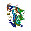
| Entry | Database: PDB / ID: 1pfz | ||||||
|---|---|---|---|---|---|---|---|
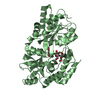
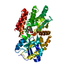
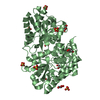
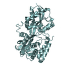
| Title | PROPLASMEPSIN II FROM PLASMODIUM FALCIPARUM | ||||||
 Components Components | PROPLASMEPSIN II | ||||||
 Keywords Keywords | ASPARTIC PROTEASE ZYMOGEN / ASPARTIC PROTEINASE ZYMOGEN / HEMOGLOBINASE / MALARIA / HYDROLASE / ASPARTYL PROTEASE / GLYCOPROTEIN | ||||||
| Function / homology |  Function and homology information Function and homology informationcytostome / plasmepsin II / acquisition of nutrients from host / vacuolar lumen / food vacuole / vacuolar membrane / aspartic-type endopeptidase activity / proteolysis Similarity search - Function | ||||||
| Biological species |  | ||||||
| Method |  X-RAY DIFFRACTION / SYNCHROTRON / MOLECULAR REPLACEMENT / Resolution: 1.85 Å X-RAY DIFFRACTION / SYNCHROTRON / MOLECULAR REPLACEMENT / Resolution: 1.85 Å | ||||||
 Authors Authors | Bernstein, N.K. / Cherney, M.M. / Loetscher, H. / Ridley, R.G. / James, M.N.G. | ||||||
 Citation Citation | Journal: Nat.Struct.Biol. / Year: 1999 Title: Crystal structure of the novel aspartic proteinase zymogen proplasmepsin II from plasmodium falciparum. Authors: Bernstein, N.K. / Cherney, M.M. / Loetscher, H. / Ridley, R.G. / James, M.N. #1: Journal: FEBS Lett. / Year: 1994Title: High Level Expression and Characterisation of Plasmepsin II, an Aspartic Proteinase from Plasmodium Falciparum Authors: Hill, J. / Tyas, L. / Phylip, L.H. / Kay, J. / Dunn, B.M. / Berry, C. | ||||||
| History |
|
- Structure visualization
| Structure viewer | Molecule: MolmilJmol/JSmol |
|---|
- Downloads & links
-Download
| PDBx/mmCIF format | 1pfz.cif.gz | 312.8 KB | Display | PDBx/mmCIF format |
|---|---|---|---|---|
| PDB format | pdb1pfz.ent.gz | 252.7 KB | Display | PDB format |
| PDBx/mmJSON format | 1pfz.json.gz | Tree view | PDBx/mmJSON format | |
| Others |  Other downloads Other downloads |
-Validation report
| Arichive directory | https://data.pdbj.org/pub/pdb/validation_reports/pf/1pfzftp://data.pdbj.org/pub/pdb/validation_reports/pf/1pfz | HTTPS FTP |
|---|
-Related structure data
| Similar structure data |
|---|


-Links
 PDBj
PDBj- Assembly
| Deposited unit | 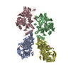
| ||||||||||||||||
|---|---|---|---|---|---|---|---|---|---|---|---|---|---|---|---|---|---|
| 1 | 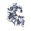
| ||||||||||||||||
| 2 | 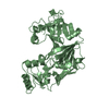
| ||||||||||||||||
| 3 | 
| ||||||||||||||||
| 4 | 
| ||||||||||||||||
| 5 | 
| ||||||||||||||||
| 6 | 
| ||||||||||||||||
| Unit cell |
| ||||||||||||||||
| Noncrystallographic symmetry (NCS) | NCS oper:
|
-Components
| #1: Protein | Mass: 42733.336 Da / Num. of mol.: 4 Mutation: 76 RESIDUES AT THE N-TERMINUS OF THE PROSEGMENT (1P - 76P) HAVE BEEN TRUNCATED, 3 RESIDUES (GLY-ARG-GLY) HAVE BEEN ADDED AT THE N-TERMINUS AS AN ARTIFACT OF THE EXPRESSION SYSTEM Source method: isolated from a genetically manipulated source Source: (gene. exp.) Cell line: BL21 / Organelle: DIGESTIVE VACUOLE / Plasmid: PBLUESCRIPT KS+ / Production host:  #2: Chemical | ChemComp-GOL /   Mass: 92.094 Da / Num. of mol.: 6 / Source method: obtained synthetically / Formula: C3H8O3 Mass: 92.094 Da / Num. of mol.: 6 / Source method: obtained synthetically / Formula: C3H8O3#3: Water | ChemComp-HOH / |  Mass: 18.015 Da / Num. of mol.: 731 / Source method: isolated from a natural source / Formula: H2O Mass: 18.015 Da / Num. of mol.: 731 / Source method: isolated from a natural source / Formula: H2OHas protein modification | Y | Sequence details | NOMENCLATURE: THE PROSEGMENT RESIDUES HAVE INSERTION CODE P (78/79 P - 118/124 P). NUMBERING FOR ...NOMENCLATU | |
|---|
-Experimental details
-Experiment
| Experiment | Method: X-RAY DIFFRACTION / Number of used crystals: 1 |
|---|
- Sample preparation
| Crystal | Density Matthews: 2.5 Å3/Da / Density % sol: 40 % | ||||||||||||||||||||||||||||||
|---|---|---|---|---|---|---|---|---|---|---|---|---|---|---|---|---|---|---|---|---|---|---|---|---|---|---|---|---|---|---|---|
| Crystal grow | pH: 7.5 / Details: pH 7.5 | ||||||||||||||||||||||||||||||
| Crystal grow | *PLUS Temperature: 105 K / Method: vapor diffusion, sitting dropDetails: 2 microlitter of protein splution was mixed with 7 microlitter of reservoir solution PH range low: 8 / PH range high: 7.5 | ||||||||||||||||||||||||||||||
| Components of the solutions | *PLUS
|
-Data collection
| Diffraction | Mean temperature: 100 K |
|---|---|
| Diffraction source | Source: SYNCHROTRON / Site: NSLS  / Beamline: X12C / Wavelength: 1 / Beamline: X12C / Wavelength: 1 |
| Detector | Type: MARRESEARCH / Detector: IMAGE PLATE AREA DETECTOR / Date: Jul 1, 1996 |
| Radiation | Monochromatic (M) / Laue (L): M / Scattering type: x-ray |
| Radiation wavelength | Wavelength: 1 Å / Relative weight: 1 |
| Reflection | Resolution: 1.95→20 Å / Num. obs: 114943 / % possible obs: 96.9 % / Redundancy: 5.3 % / Biso Wilson estimate: 10.2 Å2 / Rmerge(I) obs: 0.077 / Net I/σ(I): 7.8 |
| Reflection shell | Resolution: 1.95→2.02 Å / Redundancy: 2.5 % / Rmerge(I) obs: 0.396 / Mean I/σ(I) obs: 3 / % possible all: 91.5 |
| Reflection shell | *PLUS % possible obs: 91.5 % |
- Processing
| Software |
| ||||||||||||||||||||||||||||||||||||||||||||||||||||||||||||||||||||||||||||||||
|---|---|---|---|---|---|---|---|---|---|---|---|---|---|---|---|---|---|---|---|---|---|---|---|---|---|---|---|---|---|---|---|---|---|---|---|---|---|---|---|---|---|---|---|---|---|---|---|---|---|---|---|---|---|---|---|---|---|---|---|---|---|---|---|---|---|---|---|---|---|---|---|---|---|---|---|---|---|---|---|---|---|
| Refinement | Method to determine structure: MOLECULAR REPLACEMENT / Resolution: 1.85→20 Å / Rfactor Rfree error: 0.005 / Data cutoff high absF: 2505951.19 / Isotropic thermal model: RESTRAINED / Cross valid method: THROUGHOUT / σ(F): 0 Details: BULK SOLVENT MODEL USED DATA CUTOFF HIGH (ABS(F)) : 2505951.19 DATA CUTOFF LOW (ABS(F)) : 0.000000
| ||||||||||||||||||||||||||||||||||||||||||||||||||||||||||||||||||||||||||||||||
| Solvent computation | Solvent model: FLAT MODEL | ||||||||||||||||||||||||||||||||||||||||||||||||||||||||||||||||||||||||||||||||
| Displacement parameters | Biso mean: 23 Å2
| ||||||||||||||||||||||||||||||||||||||||||||||||||||||||||||||||||||||||||||||||
| Refine analyze |
| ||||||||||||||||||||||||||||||||||||||||||||||||||||||||||||||||||||||||||||||||
| Refinement step | Cycle: LAST / Resolution: 1.85→20 Å
| ||||||||||||||||||||||||||||||||||||||||||||||||||||||||||||||||||||||||||||||||
| Refine LS restraints |
| ||||||||||||||||||||||||||||||||||||||||||||||||||||||||||||||||||||||||||||||||
| LS refinement shell | Resolution: 1.85→1.97 Å / Rfactor Rfree error: 0.015 / Total num. of bins used: 6
| ||||||||||||||||||||||||||||||||||||||||||||||||||||||||||||||||||||||||||||||||
| Xplor file |
| ||||||||||||||||||||||||||||||||||||||||||||||||||||||||||||||||||||||||||||||||
| Software | *PLUS Name: CNS / Version: 0.3 / Classification: refinement | ||||||||||||||||||||||||||||||||||||||||||||||||||||||||||||||||||||||||||||||||
| Refinement | *PLUS | ||||||||||||||||||||||||||||||||||||||||||||||||||||||||||||||||||||||||||||||||
| Solvent computation | *PLUS | ||||||||||||||||||||||||||||||||||||||||||||||||||||||||||||||||||||||||||||||||
| Displacement parameters | *PLUS | ||||||||||||||||||||||||||||||||||||||||||||||||||||||||||||||||||||||||||||||||
| Refine LS restraints | *PLUS
| ||||||||||||||||||||||||||||||||||||||||||||||||||||||||||||||||||||||||||||||||
| LS refinement shell | *PLUS Rfactor obs: 0.276 |
