 Movie
Movie Controller

[English] 日本語
 Yorodumi
- PDB-2akc: Crystal structure of tungstate complex of the PhoN protein from S... -
+ Open data

- Basic information
| Entry | Database: PDB / ID: 2akc | ||||||
|---|---|---|---|---|---|---|---|
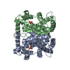
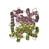
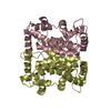
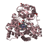
| Title | Crystal structure of tungstate complex of the PhoN protein from S. typhimurium | ||||||
 Components Components | class A nonspecific acid phosphatase PhoN | ||||||
 Keywords Keywords | HYDROLASE / Class-A bacterial non-specific acid phosphatase / tungstate complex of PhoN protein | ||||||
| Function / homology |  Function and homology information Function and homology informationacid phosphatase / acid phosphatase activity / outer membrane-bounded periplasmic space Similarity search - Function | ||||||
| Biological species |  Salmonella typhimurium (bacteria) Salmonella typhimurium (bacteria) | ||||||
| Method |  X-RAY DIFFRACTION / FOURIER SYNTHESIS / Resolution: 2.3 Å X-RAY DIFFRACTION / FOURIER SYNTHESIS / Resolution: 2.3 Å | ||||||
 Authors Authors | Makde, R.D. / Kumar, V. | ||||||
 Citation Citation | Journal: Biochemistry / Year: 2007 Title: Structure and Mutational Analysis of the PhoN Protein of Salmonella typhimurium Provide Insight into Mechanistic Details. Authors: Makde, R.D. / Mahajan, S.K. / Kumar, V. #1: Journal: Acta Crystallogr.,Sect.D / Year: 2003 Title: Purification, crystallization and preliminary x-ray diffraction studies of recombinant class A non-specific acid phosphatase of Salmonella typhimurium. Authors: Makde, R.D. / Kumar, V. / Rao, A.S. / Yadava, V.S. / Mahajan, S.K. | ||||||
| History |
|
- Structure visualization
| Structure viewer | Molecule: MolmilJmol/JSmol |
|---|
- Downloads & links
-Download
| PDBx/mmCIF format | 2akc.cif.gz | 186.5 KB | Display | PDBx/mmCIF format |
|---|---|---|---|---|
| PDB format | pdb2akc.ent.gz | 149.7 KB | Display | PDB format |
| PDBx/mmJSON format | 2akc.json.gz | Tree view | PDBx/mmJSON format | |
| Others |  Other downloads Other downloads |
-Validation report
| Arichive directory | https://data.pdbj.org/pub/pdb/validation_reports/ak/2akcftp://data.pdbj.org/pub/pdb/validation_reports/ak/2akc | HTTPS FTP |
|---|
-Related structure data
| Related structure data |  2a96SC  2ipbC S: Starting model for refinement C: citing same article ( |
|---|---|
| Similar structure data |


-Links
 PDBj
PDBj
- Assembly
| Deposited unit | 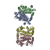
| ||||||||
|---|---|---|---|---|---|---|---|---|---|
| 1 | 
| ||||||||
| 2 | 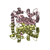
| ||||||||
| Unit cell |
| ||||||||
| Details | The entry contains crystallographic asymmetric unit consisting of four chains (two dimers). A dimer is the known biologically active state of the PHON protein. Chains A and B form one dimer while the chains C and D form the other dimer. |
-Components
| #1: Protein | Mass: 26090.242 Da / Num. of mol.: 4 / Fragment: Periplasmic (mature) PhoN (residues 21-250) Source method: isolated from a genetically manipulated source Source: (gene. exp.) Salmonella typhimurium (bacteria) / Gene: phoN / Plasmid: pSK- / Production host: References: GenBank: 21913311, UniProt: Q8KRU6*PLUS, acid phosphatase #2: Chemical | ChemComp-WO4 /   Mass: 247.838 Da / Num. of mol.: 4 / Source method: obtained synthetically / Formula: WO4 Mass: 247.838 Da / Num. of mol.: 4 / Source method: obtained synthetically / Formula: WO4#3: Water | ChemComp-HOH / |  Mass: 18.015 Da / Num. of mol.: 326 / Source method: isolated from a natural source / Formula: H2O Mass: 18.015 Da / Num. of mol.: 326 / Source method: isolated from a natural source / Formula: H2OHas protein modification | Y | |
|---|
-Experimental details
-Experiment
| Experiment | Method: X-RAY DIFFRACTION / Number of used crystals: 1 |
|---|
- Sample preparation
| Crystal | Density Matthews: 2.16 Å3/Da / Density % sol: 43 % |
|---|---|
| Crystal grow | Temperature: 293 K / Method: vapor diffusion, hanging drop / pH: 5.6 Details: PEG-6000 15%, Glycerol 2%, 1mM sodium tungstate, 25mM sodium acetate, pH 5.6, VAPOR DIFFUSION, HANGING DROP, temperature 293K |
-Data collection
| Diffraction | Mean temperature: 293 K |
|---|---|
| Diffraction source | Source: ROTATING ANODE / Type: RIGAKU RU200 / Wavelength: 1.5418 Å |
| Detector | Type: MARRESEARCH / Detector: IMAGE PLATE / Date: Aug 3, 2002 / Details: OSMIC MIRRORS |
| Radiation | Monochromator: Ni filter / Protocol: SINGLE WAVELENGTH / Monochromatic (M) / Laue (L): M / Scattering type: x-ray |
| Radiation wavelength | Wavelength: 1.5418 Å / Relative weight: 1 |
| Reflection | Resolution: 2.3→30 Å / Num. all: 37751 / Num. obs: 37069 / % possible obs: 93 % / Observed criterion σ(I): 0 / Redundancy: 1.8 % / Biso Wilson estimate: 30.34 Å2 / Rmerge(I) obs: 0.06 / Net I/σ(I): 9.8 |
| Reflection shell | Resolution: 2.3→2.42 Å / Redundancy: 1.7 % / Rmerge(I) obs: 0.11 / Mean I/σ(I) obs: 3.2 / Num. unique all: 5148 / % possible all: 89.2 |
- Processing
| Software |
| ||||||||||||||||||||||||||||||||||||
|---|---|---|---|---|---|---|---|---|---|---|---|---|---|---|---|---|---|---|---|---|---|---|---|---|---|---|---|---|---|---|---|---|---|---|---|---|---|
| Refinement | Method to determine structure: FOURIER SYNTHESIS Starting model: PDB entry 2A96 Resolution: 2.3→20 Å / Isotropic thermal model: ANISOTROPIC / Cross valid method: THROUGHOUT / σ(F): 0 / Stereochemistry target values: Engh & Huber Details: The submission is the structure of periplasmic (mature) PhoN protein constituted of 230 amino acid residues. The 20 N-terminus signal peptide residues corresponding to the GenBank entry ...Details: The submission is the structure of periplasmic (mature) PhoN protein constituted of 230 amino acid residues. The 20 N-terminus signal peptide residues corresponding to the GenBank entry 21913311 (AAM81208) are known to be cleaved and thus not observed in the mature protein. The residue numbering in the deposited structure however is as the full length CDS in the Genbank entry.
| ||||||||||||||||||||||||||||||||||||
| Solvent computation | Solvent model: Bulk Solvent modeling, Flat model / Bsol: 39.6 Å2 / ksol: 0.328 e/Å3 | ||||||||||||||||||||||||||||||||||||
| Displacement parameters | Biso mean: 28.9 Å2
| ||||||||||||||||||||||||||||||||||||
| Refine analyze |
| ||||||||||||||||||||||||||||||||||||
| Refinement step | Cycle: LAST / Resolution: 2.3→20 Å
| ||||||||||||||||||||||||||||||||||||
| Refine LS restraints |
| ||||||||||||||||||||||||||||||||||||
| LS refinement shell | Resolution: 2.3→2.38 Å / Total num. of bins used: 10
| ||||||||||||||||||||||||||||||||||||
| Xplor file |
|
