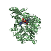
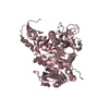
Deposited unit
A: AMP-dependent synthetase and ligase
B: AMP-dependent synthetase and ligase
C: AMP-dependent synthetase and ligase
D: AMP-dependent synthetase and ligase
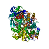
hetero molecules Summary Component details
Theoretical mass Number of molelcules Total (without water) 238,291 12 Polymers 236,420 4 Non-polymers 1,871 8 Water 4,450 247
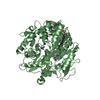
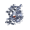
1
A: AMP-dependent synthetase and ligase
hetero molecules Summary Component details Symmetry operations Calculated values
Theoretical mass Number of molelcules Total (without water) 59,573 3 Polymers 59,105 1 Non-polymers 468 2 Water 18 1
Type Name Symmetry operation Number identity operation 1_555 x,y,z 1
Buried area 120 Å2 ΔGint -10 kcal/mol Surface area 21990 Å2 Method
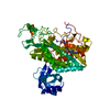
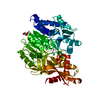
2
B: AMP-dependent synthetase and ligase
hetero molecules Summary Component details Symmetry operations Calculated values
Theoretical mass Number of molelcules Total (without water) 59,573 3 Polymers 59,105 1 Non-polymers 468 2 Water 18 1
Type Name Symmetry operation Number identity operation 1_555 x,y,z 1
Buried area 120 Å2 ΔGint -10 kcal/mol Surface area 21630 Å2 Method
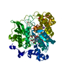
3
C: AMP-dependent synthetase and ligase
hetero molecules Summary Component details Symmetry operations Calculated values
Theoretical mass Number of molelcules Total (without water) 59,573 3 Polymers 59,105 1 Non-polymers 468 2 Water 18 1
Type Name Symmetry operation Number identity operation 1_555 x,y,z 1
Buried area 120 Å2 ΔGint -10 kcal/mol Surface area 22170 Å2 Method
4
D: AMP-dependent synthetase and ligase
hetero molecules Summary Component details Symmetry operations Calculated values
Theoretical mass Number of molelcules Total (without water) 59,573 3 Polymers 59,105 1 Non-polymers 468 2 Water 18 1
Type Name Symmetry operation Number identity operation 1_555 x,y,z 1
Buried area 120 Å2 ΔGint -11 kcal/mol Surface area 21620 Å2 Method
Unit cell Length a, b, c (Å) 65.750, 104.450, 120.000 Angle α, β, γ (deg.) 81.78, 83.01, 81.98 Int Tables number 1 Space group name H-M P1
Noncrystallographic symmetry (NCS) NCS domain ID Ens-ID Details (eV)1 1 A2 1 B1 2 A2 2 C1 3 A2 3 D1 4 B2 4 C1 5 B2 5 D1 6 C2 6 D
NCS domain segments Component-ID / End auth comp-ID / End label comp-ID / Refine code
Dom-ID Ens-ID Beg auth comp-ID Beg label comp-ID Auth asym-ID Label asym-ID Auth seq-ID Label seq-ID 1 1 ALAALAAA9 - 508 29 - 528 2 1 ALAALABB9 - 508 29 - 528 1 2 ALAALAAA9 - 508 29 - 528 2 2 ALAALACC9 - 508 29 - 528 1 3 ALAALAAA9 - 508 29 - 528 2 3 ALAALADD9 - 508 29 - 528 1 4 ALAALABB9 - 508 29 - 528 2 4 ALAALACC9 - 508 29 - 528 1 5 VALVALBB5 - 508 25 - 528 2 5 VALVALDD5 - 508 25 - 528 1 6 ALAALACC9 - 508 29 - 528 2 6 ALAALADD9 - 508 29 - 528
NCS ensembles
 Movie
Movie Controller


 Open data
 Basic information
 Components
 Keywords
 Function and homology information
 Streptomyces sp. ML694-90F3 (bacteria)
 X-RAY DIFFRACTION /
  Authors
 Japan, 1items
  Citation
 Structure visualization
 Downloads & links
 Other downloads
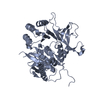


 PDBj
PDBj
 Assembly


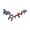
 Mass: 432.326 Da / Num. of mol.: 4 / Source method: obtained synthetically / Formula: C14H21N6O8P
 Mass: 35.453 Da / Num. of mol.: 4 / Source method: obtained synthetically / Formula: Cl
 Mass: 18.015 Da / Num. of mol.: 247 / Source method: isolated from a natural source / Formula: H2O
 Sample preparation
 Processing