 Movie
Movie Controller

[English] 日本語
 Yorodumi
- PDB-3eq6: Crystal structure of human acyl-CoA synthetase medium-chain famil... -
+ Open data

- Basic information
| Entry | Database: PDB / ID: 3eq6 | ||||||
|---|---|---|---|---|---|---|---|
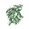
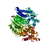
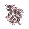
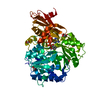
| Title | Crystal structure of human acyl-CoA synthetase medium-chain family member 2A (L64P mutation) in a ternary complex with products | ||||||
 Components Components | Acyl-coenzyme A synthetase ACSM2A | ||||||
 Keywords Keywords | LIGASE / Middle-chain acyl-CoA synthetase / xenobiotic/medium-chain fatty acid-CoA ligase / ATP-binding / fatty acid metabolism / lipid metabolism / magnesium / metal-binding / mitochondrion / nucleotide-binding polymorphism / transit peptide / Structural Genomics / SGC / Structural Genomics Consortium | ||||||
| Function / homology |  Function and homology information Function and homology informationdecanoate-CoA ligase activity / Conjugation of salicylate with glycine / medium-chain acyl-CoA ligase / fatty acid ligase activity / benzoate-CoA ligase / benzoate-CoA ligase activity / medium-chain fatty-acyl-CoA metabolic process / medium-chain fatty acid-CoA ligase activity / fatty-acyl-CoA synthase activity / acyl-CoA metabolic process ...decanoate-CoA ligase activity / Conjugation of salicylate with glycine / medium-chain acyl-CoA ligase / fatty acid ligase activity / benzoate-CoA ligase / benzoate-CoA ligase activity / medium-chain fatty-acyl-CoA metabolic process / medium-chain fatty acid-CoA ligase activity / fatty-acyl-CoA synthase activity / acyl-CoA metabolic process / triglyceride homeostasis / Aspirin ADME / fatty acid biosynthetic process / glucose homeostasis / mitochondrial matrix / mitochondrion / ATP binding / metal ion binding Similarity search - Function | ||||||
| Biological species |  Homo sapiens (human) Homo sapiens (human) | ||||||
| Method |  X-RAY DIFFRACTION / MOLECULAR REPLACEMENT / Resolution: 2.4 Å X-RAY DIFFRACTION / MOLECULAR REPLACEMENT / Resolution: 2.4 Å | ||||||
 Authors Authors | Pilka, E.S. / Kochan, G. / Yue, W.W. / Bhatia, C. / Von delft, F. / Arrowsmith, C.H. / Edwards, A.M. / Weigelt, J. / Bountra, C. / Oppermann, U. / Structural Genomics Consortium (SGC) | ||||||
 Citation Citation | Journal: J.Mol.Biol. / Year: 2009 Title: Structural snapshots for the conformation-dependent catalysis by human medium-chain acyl-coenzyme A synthetase ACSM2A Authors: Kochan, G. / Pilka, E.S. / von Delft, F. / Oppermann, U. / Yue, W.W. | ||||||
| History |
|
- Structure visualization
| Structure viewer | Molecule: MolmilJmol/JSmol |
|---|
- Downloads & links
-Download
| PDBx/mmCIF format | 3eq6.cif.gz | 234.4 KB | Display | PDBx/mmCIF format |
|---|---|---|---|---|
| PDB format | pdb3eq6.ent.gz | 183.3 KB | Display | PDB format |
| PDBx/mmJSON format | 3eq6.json.gz | Tree view | PDBx/mmJSON format | |
| Others |  Other downloads Other downloads |
-Validation report
| Arichive directory | https://data.pdbj.org/pub/pdb/validation_reports/eq/3eq6ftp://data.pdbj.org/pub/pdb/validation_reports/eq/3eq6 | HTTPS FTP |
|---|
-Related structure data
| Related structure data | 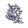 2vzeC 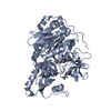 2wd9C 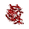 3b7wSC 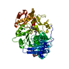 3c5eC  3dayC 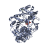 3gpcC C: citing same article ( S: Starting model for refinement |
|---|---|
| Similar structure data |


-Links
 PDBj
PDBj


- Assembly
| Deposited unit | 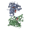
| ||||||||||||||||||
|---|---|---|---|---|---|---|---|---|---|---|---|---|---|---|---|---|---|---|---|
| 1 | 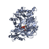
| ||||||||||||||||||
| 2 | 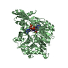
| ||||||||||||||||||
| Unit cell |
| ||||||||||||||||||
| Noncrystallographic symmetry (NCS) | NCS domain:
NCS domain segments: Component-ID: 1 / Ens-ID: 1 / Beg auth comp-ID: HIS / Beg label comp-ID: HIS / End auth comp-ID: LYS / End label comp-ID: LYS / Refine code: 3 / Auth seq-ID: 37 - 569 / Label seq-ID: 30 - 562
|
-Components
| #1: Protein | Mass: 63334.562 Da / Num. of mol.: 2 / Fragment: UNP residues 32-577 / Mutation: L64P Source method: isolated from a genetically manipulated source Source: (gene. exp.) Homo sapiens (human) / Gene: ACSM2A / Production host:  Trichoplusia ni (cabbage looper) / Strain (production host): high five / References: UniProt: Q08AH3, medium-chain acyl-CoA ligase Trichoplusia ni (cabbage looper) / Strain (production host): high five / References: UniProt: Q08AH3, medium-chain acyl-CoA ligase#2: Chemical |   Mass: 347.221 Da / Num. of mol.: 2 / Source method: obtained synthetically / Formula: C10H14N5O7P / Comment: AMP*YM Mass: 347.221 Da / Num. of mol.: 2 / Source method: obtained synthetically / Formula: C10H14N5O7P / Comment: AMP*YM#3: Chemical |   Mass: 837.624 Da / Num. of mol.: 2 / Source method: obtained synthetically / Formula: C25H42N7O17P3S Mass: 837.624 Da / Num. of mol.: 2 / Source method: obtained synthetically / Formula: C25H42N7O17P3S#4: Water | ChemComp-HOH / |  Mass: 18.015 Da / Num. of mol.: 447 / Source method: isolated from a natural source / Formula: H2O Mass: 18.015 Da / Num. of mol.: 447 / Source method: isolated from a natural source / Formula: H2O |
|---|
-Experimental details
-Experiment
| Experiment | Method: X-RAY DIFFRACTION / Number of used crystals: 1 |
|---|
- Sample preparation
| Crystal | Density Matthews: 2.57 Å3/Da / Density % sol: 52.1 % |
|---|---|
| Crystal grow | Temperature: 293 K / Method: vapor diffusion, hanging drop / pH: 7.5 Details: 0.1M Mg(form), 15% PEG3350, pH7.5, VAPOR DIFFUSION, HANGING DROP, temperature 293K |
-Data collection
| Diffraction | Mean temperature: 100 K |
|---|---|
| Diffraction source | Source: ROTATING ANODE / Type: RIGAKU FR-E SUPERBRIGHT / Wavelength: 1.5418 Å |
| Detector | Type: RIGAKU RAXIS HTC / Detector: IMAGE PLATE / Date: Jul 4, 2008 / Details: OSMIC |
| Radiation | Monochromator: NI / Protocol: SINGLE WAVELENGTH / Monochromatic (M) / Laue (L): M / Scattering type: x-ray |
| Radiation wavelength | Wavelength: 1.5418 Å / Relative weight: 1 |
| Reflection | Resolution: 2.4→24.99 Å / Num. all: 49866 / Num. obs: 47300 / % possible obs: 99.3 % / Observed criterion σ(F): 2 / Observed criterion σ(I): 2 / Redundancy: 3.3 % / Rmerge(I) obs: 0.107 / Rsym value: 0.07 / Net I/σ(I): 8 |
| Reflection shell | Resolution: 2.4→2.53 Å / Redundancy: 3.1 % / Rmerge(I) obs: 0.494 / Mean I/σ(I) obs: 2.2 / Num. unique all: 7167 / Rsym value: 0.323 / % possible all: 98 |
- Processing
| Software |
| ||||||||||||||||||||||||||||||||||||||||||||||||||||||||||||||||||||||||||||||||||||||||||||||||||||||||||||||||||||||||||||||||||||||||||||||||||||||||||||||||||||||||||
|---|---|---|---|---|---|---|---|---|---|---|---|---|---|---|---|---|---|---|---|---|---|---|---|---|---|---|---|---|---|---|---|---|---|---|---|---|---|---|---|---|---|---|---|---|---|---|---|---|---|---|---|---|---|---|---|---|---|---|---|---|---|---|---|---|---|---|---|---|---|---|---|---|---|---|---|---|---|---|---|---|---|---|---|---|---|---|---|---|---|---|---|---|---|---|---|---|---|---|---|---|---|---|---|---|---|---|---|---|---|---|---|---|---|---|---|---|---|---|---|---|---|---|---|---|---|---|---|---|---|---|---|---|---|---|---|---|---|---|---|---|---|---|---|---|---|---|---|---|---|---|---|---|---|---|---|---|---|---|---|---|---|---|---|---|---|---|---|---|---|---|---|
| Refinement | Method to determine structure: MOLECULAR REPLACEMENT Starting model: PDB ENTRY 3B7W Resolution: 2.4→21.73 Å / Cor.coef. Fo:Fc: 0.94 / Cor.coef. Fo:Fc free: 0.909 / SU B: 15.136 / SU ML: 0.161 / TLS residual ADP flag: LIKELY RESIDUAL / Cross valid method: THROUGHOUT / σ(F): 0 / σ(I): 0 / ESU R: 0.394 / ESU R Free: 0.25 / Stereochemistry target values: MAXIMUM LIKELIHOOD / Details: HYDROGENS HAVE BEEN ADDED IN THE RIDING POSITIONS
| ||||||||||||||||||||||||||||||||||||||||||||||||||||||||||||||||||||||||||||||||||||||||||||||||||||||||||||||||||||||||||||||||||||||||||||||||||||||||||||||||||||||||||
| Solvent computation | Ion probe radii: 0.8 Å / Shrinkage radii: 0.8 Å / VDW probe radii: 1 Å / Solvent model: MASK | ||||||||||||||||||||||||||||||||||||||||||||||||||||||||||||||||||||||||||||||||||||||||||||||||||||||||||||||||||||||||||||||||||||||||||||||||||||||||||||||||||||||||||
| Displacement parameters | Biso mean: 17.19 Å2
| ||||||||||||||||||||||||||||||||||||||||||||||||||||||||||||||||||||||||||||||||||||||||||||||||||||||||||||||||||||||||||||||||||||||||||||||||||||||||||||||||||||||||||
| Refinement step | Cycle: LAST / Resolution: 2.4→21.73 Å
| ||||||||||||||||||||||||||||||||||||||||||||||||||||||||||||||||||||||||||||||||||||||||||||||||||||||||||||||||||||||||||||||||||||||||||||||||||||||||||||||||||||||||||
| Refine LS restraints |
| ||||||||||||||||||||||||||||||||||||||||||||||||||||||||||||||||||||||||||||||||||||||||||||||||||||||||||||||||||||||||||||||||||||||||||||||||||||||||||||||||||||||||||
| Refine LS restraints NCS | Dom-ID: 1 / Auth asym-ID: A / Ens-ID: 1 / Refine-ID: X-RAY DIFFRACTION
| ||||||||||||||||||||||||||||||||||||||||||||||||||||||||||||||||||||||||||||||||||||||||||||||||||||||||||||||||||||||||||||||||||||||||||||||||||||||||||||||||||||||||||
| LS refinement shell | Resolution: 2.4→2.462 Å / Total num. of bins used: 20
| ||||||||||||||||||||||||||||||||||||||||||||||||||||||||||||||||||||||||||||||||||||||||||||||||||||||||||||||||||||||||||||||||||||||||||||||||||||||||||||||||||||||||||
| Refinement TLS params. | Method: refined / Refine-ID: X-RAY DIFFRACTION
| ||||||||||||||||||||||||||||||||||||||||||||||||||||||||||||||||||||||||||||||||||||||||||||||||||||||||||||||||||||||||||||||||||||||||||||||||||||||||||||||||||||||||||
| Refinement TLS group |
|
