 Movie
Movie Controller

[English] 日本語
 Yorodumi
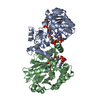
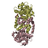
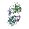
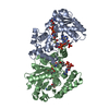
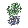
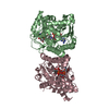
- PDB-5j3h: Human insulin receptor domains L1-CR in complex with peptide S519... -
+ Open data

- Basic information
| Entry | Database: PDB / ID: 5j3h | |||||||||||||||
|---|---|---|---|---|---|---|---|---|---|---|---|---|---|---|---|---|
| Title | Human insulin receptor domains L1-CR in complex with peptide S519C16 and 83-7 Fv | |||||||||||||||
 Components Components |
| |||||||||||||||
 Keywords Keywords | HORMONE RECEPTOR/IMMUNE SYSTEM / Insulin receptor Insulin mimetic peptide Fv fragment / HORMONE RECEPTOR-IMMUNE SYSTEM complex | |||||||||||||||
| Function / homology |  Function and homology information Function and homology informationregulation of female gonad development / positive regulation of meiotic cell cycle / insulin-like growth factor II binding / positive regulation of developmental growth / male sex determination / insulin receptor complex / insulin-like growth factor I binding / insulin receptor activity / positive regulation of protein-containing complex disassembly / exocrine pancreas development ...regulation of female gonad development / positive regulation of meiotic cell cycle / insulin-like growth factor II binding / positive regulation of developmental growth / male sex determination / insulin receptor complex / insulin-like growth factor I binding / insulin receptor activity / positive regulation of protein-containing complex disassembly / exocrine pancreas development / dendritic spine maintenance / insulin binding / adrenal gland development / cargo receptor activity / PTB domain binding / Signaling by Insulin receptor / IRS activation / neuronal cell body membrane / positive regulation of respiratory burst / amyloid-beta clearance / regulation of embryonic development / insulin receptor substrate binding / positive regulation of receptor internalization / epidermis development / positive regulation of glycogen biosynthetic process / Signal attenuation / protein kinase activator activity / transport across blood-brain barrier / phosphatidylinositol 3-kinase binding / heart morphogenesis / Insulin receptor recycling / insulin-like growth factor receptor binding / neuron projection maintenance / positive regulation of mitotic nuclear division / Insulin receptor signalling cascade / receptor-mediated endocytosis / dendrite membrane / positive regulation of glycolytic process / positive regulation of D-glucose import across plasma membrane / learning / receptor protein-tyrosine kinase / caveola / receptor internalization / male gonad development / cellular response to growth factor stimulus / cellular response to insulin stimulus / memory / positive regulation of nitric oxide biosynthetic process / insulin receptor signaling pathway / protein autophosphorylation / late endosome / glucose homeostasis / amyloid-beta binding / PI5P, PP2A and IER3 Regulate PI3K/AKT Signaling / protein tyrosine kinase activity / positive regulation of canonical NF-kappaB signal transduction / positive regulation of MAPK cascade / lysosome / positive regulation of phosphatidylinositol 3-kinase/protein kinase B signal transduction / signaling receptor complex / endosome membrane / positive regulation of cell migration / G protein-coupled receptor signaling pathway / external side of plasma membrane / protein domain specific binding / axon / positive regulation of cell population proliferation / symbiont entry into host cell / regulation of DNA-templated transcription / positive regulation of DNA-templated transcription / GTP binding / protein-containing complex binding / extracellular exosome / ATP binding / membrane / identical protein binding / plasma membrane Similarity search - Function | |||||||||||||||
| Biological species |   Homo sapiens (human) Homo sapiens (human) Endothia gyrosa (fungus) Endothia gyrosa (fungus) | |||||||||||||||
| Method |  X-RAY DIFFRACTION / SYNCHROTRON / MOLECULAR REPLACEMENT / Resolution: 3.27 Å X-RAY DIFFRACTION / SYNCHROTRON / MOLECULAR REPLACEMENT / Resolution: 3.27 Å | |||||||||||||||
 Authors Authors | Lawrence, M. / Menting, J. / Lawrence, C. | |||||||||||||||
| Funding support |  Australia, 4items Australia, 4items
| |||||||||||||||
 Citation Citation | Journal: J.Biol.Chem. / Year: 2016 Title: Insulin Mimetic Peptide Disrupts the Primary Binding Site of the Insulin Receptor. Authors: Lawrence, C.F. / Margetts, M.B. / Menting, J.G. / Smith, N.A. / Smith, B.J. / Ward, C.W. / Lawrence, M.C. | |||||||||||||||
| History |
|
- Structure visualization
| Structure viewer | Molecule: MolmilJmol/JSmol |
|---|
- Downloads & links
-Download
| PDBx/mmCIF format | 5j3h.cif.gz | 229.4 KB | Display | PDBx/mmCIF format |
|---|---|---|---|---|
| PDB format | pdb5j3h.ent.gz | 184 KB | Display | PDB format |
| PDBx/mmJSON format | 5j3h.json.gz | Tree view | PDBx/mmJSON format | |
| Others |  Other downloads Other downloads |
-Validation report
| Arichive directory | https://data.pdbj.org/pub/pdb/validation_reports/j3/5j3hftp://data.pdbj.org/pub/pdb/validation_reports/j3/5j3h | HTTPS FTP |
|---|
-Related structure data
| Related structure data | 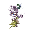 4ogaS S: Starting model for refinement |
|---|---|
| Similar structure data |


-Links
 PDBj
PDBj


- Assembly
| Deposited unit | 
| ||||||||
|---|---|---|---|---|---|---|---|---|---|
| 1 |
| ||||||||
| Unit cell |
|
-Components
-Antibody , 2 types, 2 molecules CD
| #2: Antibody | Mass: 13440.069 Da / Num. of mol.: 1 Source method: isolated from a genetically manipulated source Source: (gene. exp.)  Brevibacillus (bacteria) Brevibacillus (bacteria) |
|---|---|
| #3: Antibody | Mass: 13152.705 Da / Num. of mol.: 1 Source method: isolated from a genetically manipulated source Source: (gene. exp.) Brevibacillus (bacteria) |
-Protein/peptide / Protein , 2 types, 2 molecules BE
| #1: Protein/peptide | Mass: 1950.047 Da / Num. of mol.: 1 / Source method: obtained synthetically Details: 16 C-terminal residues of peptide S519: SER-LEU-ASP-GLU-SER-PHE-TYR-ASP-TRP-PHE-GLU-ARG-GLN-LEU Source: (synth.) Endothia gyrosa (fungus) |
|---|---|
| #4: Protein | Mass: 36231.762 Da / Num. of mol.: 1 / Fragment: UNP residues 28-337 Source method: isolated from a genetically manipulated source Source: (gene. exp.) Homo sapiens (human) / Gene: INSR / Production host:  Cricetulus griseus (Chinese hamster) / Strain (production host): Lec8 Cricetulus griseus (Chinese hamster) / Strain (production host): Lec8References: UniProt: P06213, receptor protein-tyrosine kinase |
-Sugars , 4 types, 5 molecules 
| #5: Polysaccharide | 2-acetamido-2-deoxy-beta-D-glucopyranose-(1-4)-[alpha-L-fucopyranose-(1-6)]2-acetamido-2-deoxy-beta- ...2-acetamido-2-deoxy-beta-D-glucopyranose-(1-4)-[alpha-L-fucopyranose-(1-6)]2-acetamido-2-deoxy-beta-D-glucopyranose Source method: isolated from a genetically manipulated source |
|---|---|
| #6: Polysaccharide | 2-acetamido-2-deoxy-beta-D-glucopyranose-(1-4)-2-acetamido-2-deoxy-beta-D-glucopyranose Source method: isolated from a genetically manipulated source |
| #7: Polysaccharide | 2-acetamido-2-deoxy-beta-D-glucopyranose-(1-2)-alpha-D-mannopyranose-(1-3)-[alpha-D-mannopyranose- ...2-acetamido-2-deoxy-beta-D-glucopyranose-(1-2)-alpha-D-mannopyranose-(1-3)-[alpha-D-mannopyranose-(1-6)]beta-D-mannopyranose-(1-4)-2-acetamido-2-deoxy-beta-D-glucopyranose-(1-4)-[alpha-L-fucopyranose-(1-6)]2-acetamido-2-deoxy-beta-D-glucopyranose Source method: isolated from a genetically manipulated source |
| #8: Sugar |  Type: D-saccharide, beta linking / Mass: 221.208 Da / Num. of mol.: 2 Type: D-saccharide, beta linking / Mass: 221.208 Da / Num. of mol.: 2Source method: isolated from a genetically manipulated source Formula: C8H15NO6 |
-Details
| Has protein modification | Y |
|---|
-Experimental details
-Experiment
| Experiment | Method: X-RAY DIFFRACTION / Number of used crystals: 1 |
|---|
- Sample preparation
| Crystal | Density Matthews: 5.2 Å3/Da / Density % sol: 76.33 % |
|---|---|
| Crystal grow | Temperature: 300 K / Method: vapor diffusion, sitting drop / Details: 1.75 M (NH4)2HPO4 |
-Data collection
| Diffraction | Mean temperature: 100 K |
|---|---|
| Diffraction source | Source: SYNCHROTRON / Site: Australian Synchrotron / Beamline: MX2 / Wavelength: 1 Å |
| Detector | Type: ADSC QUANTUM 315r / Detector: CCD / Date: Jul 1, 2015 |
| Radiation | Protocol: SINGLE WAVELENGTH / Monochromatic (M) / Laue (L): M / Scattering type: x-ray |
| Radiation wavelength | Wavelength: 1 Å / Relative weight: 1 |
| Reflection | Resolution: 3.27→34.79 Å / Num. obs: 21483 / % possible obs: 99.4 % / Redundancy: 6.3 % / Biso Wilson estimate: 85.67 Å2 / Net I/σ(I): 7.68 |
| Reflection shell | Resolution: 3.27→3.39 Å / Redundancy: 6.38 % / Rmerge(I) obs: 2.23 / Mean I/σ(I) obs: 1 / % possible all: 98.9 |
- Processing
| Software |
| ||||||||||||||||||||||||||||||||||||||||||||||||||||||||||||||||||||||||||||||||||||||||||||||||||||||||||||||||||||||||||||||||||||||||||||||||||||||
|---|---|---|---|---|---|---|---|---|---|---|---|---|---|---|---|---|---|---|---|---|---|---|---|---|---|---|---|---|---|---|---|---|---|---|---|---|---|---|---|---|---|---|---|---|---|---|---|---|---|---|---|---|---|---|---|---|---|---|---|---|---|---|---|---|---|---|---|---|---|---|---|---|---|---|---|---|---|---|---|---|---|---|---|---|---|---|---|---|---|---|---|---|---|---|---|---|---|---|---|---|---|---|---|---|---|---|---|---|---|---|---|---|---|---|---|---|---|---|---|---|---|---|---|---|---|---|---|---|---|---|---|---|---|---|---|---|---|---|---|---|---|---|---|---|---|---|---|---|---|---|---|
| Refinement | Method to determine structure: MOLECULAR REPLACEMENT Starting model: 4OGA Resolution: 3.27→34.79 Å / Cor.coef. Fo:Fc: 0.9116 / Cor.coef. Fo:Fc free: 0.8871 / SU R Cruickshank DPI: 0.72 / Cross valid method: THROUGHOUT / σ(F): 0 / SU R Blow DPI: 0.725 / SU Rfree Blow DPI: 0.357 / SU Rfree Cruickshank DPI: 0.361
| ||||||||||||||||||||||||||||||||||||||||||||||||||||||||||||||||||||||||||||||||||||||||||||||||||||||||||||||||||||||||||||||||||||||||||||||||||||||
| Displacement parameters | Biso mean: 122.88 Å2
| ||||||||||||||||||||||||||||||||||||||||||||||||||||||||||||||||||||||||||||||||||||||||||||||||||||||||||||||||||||||||||||||||||||||||||||||||||||||
| Refine analyze | Luzzati coordinate error obs: 0.899 Å | ||||||||||||||||||||||||||||||||||||||||||||||||||||||||||||||||||||||||||||||||||||||||||||||||||||||||||||||||||||||||||||||||||||||||||||||||||||||
| Refinement step | Cycle: LAST / Resolution: 3.27→34.79 Å
| ||||||||||||||||||||||||||||||||||||||||||||||||||||||||||||||||||||||||||||||||||||||||||||||||||||||||||||||||||||||||||||||||||||||||||||||||||||||
| Refine LS restraints |
| ||||||||||||||||||||||||||||||||||||||||||||||||||||||||||||||||||||||||||||||||||||||||||||||||||||||||||||||||||||||||||||||||||||||||||||||||||||||
| LS refinement shell | Resolution: 3.27→3.43 Å / Total num. of bins used: 11
| ||||||||||||||||||||||||||||||||||||||||||||||||||||||||||||||||||||||||||||||||||||||||||||||||||||||||||||||||||||||||||||||||||||||||||||||||||||||
| Refinement TLS params. | Method: refined / Refine-ID: X-RAY DIFFRACTION
| ||||||||||||||||||||||||||||||||||||||||||||||||||||||||||||||||||||||||||||||||||||||||||||||||||||||||||||||||||||||||||||||||||||||||||||||||||||||
| Refinement TLS group |
|
