 Movie
Movie Controller

[English] 日本語
 Yorodumi
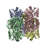
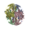
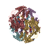
- PDB-5izd: Wild-type glyceraldehyde dehydrogenase from Thermoplasma acidophi... -
+ Open data

- Basic information
| Entry | Database: PDB / ID: 5izd | ||||||
|---|---|---|---|---|---|---|---|
| Title | Wild-type glyceraldehyde dehydrogenase from Thermoplasma acidophilum in complex with NADP | ||||||
 Components Components | D-glyceraldehyde dehydrogenase (NADP(+)) | ||||||
 Keywords Keywords | OXIDOREDUCTASE / NADP-dependent dehydrogenase | ||||||
| Function / homology |  Function and homology information Function and homology informationD-glyceraldehyde dehydrogenase (NADP+) / glyceraldehyde dehydrogenase (NADP+) activity / : / succinate-semialdehyde dehydrogenase (NAD+) activity / GABA catabolic process / glycolytic process / protein homotetramerization / protein homodimerization activity / cytosol Similarity search - Function | ||||||
| Biological species |   Thermoplasma acidophilum (acidophilic) Thermoplasma acidophilum (acidophilic) | ||||||
| Method |  X-RAY DIFFRACTION / SYNCHROTRON / MOLECULAR REPLACEMENT / Resolution: 2.05 Å X-RAY DIFFRACTION / SYNCHROTRON / MOLECULAR REPLACEMENT / Resolution: 2.05 Å | ||||||
 Authors Authors | Iermak, I. / Mesters, J.R. / Kuta Smatanova, I. | ||||||
| Funding support |  Czech Republic, 1items Czech Republic, 1items
| ||||||
 Citation Citation | Journal: To Be Published Title: Structure of wild-type glyceraldehyde dehydrogenase from Thermoplasma acidophilum in complex with NADP Authors: Iermak, I. / Mesters, J.R. / Steffler, F. / Sieber, V. / Kuta Smatanova, I. | ||||||
| History |
|
- Structure visualization
| Structure viewer | Molecule: MolmilJmol/JSmol |
|---|
- Downloads & links
-Download
| PDBx/mmCIF format | 5izd.cif.gz | 1.4 MB | Display | PDBx/mmCIF format |
|---|---|---|---|---|
| PDB format | pdb5izd.ent.gz | 1.2 MB | Display | PDB format |
| PDBx/mmJSON format | 5izd.json.gz | Tree view | PDBx/mmJSON format | |
| Others |  Other downloads Other downloads |
-Validation report
| Arichive directory | https://data.pdbj.org/pub/pdb/validation_reports/iz/5izdftp://data.pdbj.org/pub/pdb/validation_reports/iz/5izd | HTTPS FTP |
|---|
-Related structure data
| Related structure data |  5j77C 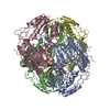 3k2wS S: Starting model for refinement C: citing same article ( |
|---|---|
| Similar structure data |


-Links
 PDBj
PDBj

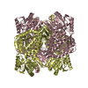
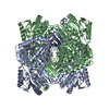
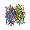
- Assembly
| Deposited unit | 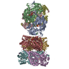
| ||||||||
|---|---|---|---|---|---|---|---|---|---|
| 1 | 
| ||||||||
| 2 | 
| ||||||||
| Unit cell |
|
-Components
| #1: Protein | Mass: 56548.973 Da / Num. of mol.: 8 Source method: isolated from a genetically manipulated source Source: (gene. exp.) Thermoplasma acidophilum (acidophilic) / Gene: Ta0809 / Plasmid: pCBRHisC / Production host:  References: UniProt: Q9HK01, D-glyceraldehyde dehydrogenase (NADP+) #2: Chemical | ChemComp-NAP /   Mass: 743.405 Da / Num. of mol.: 6 / Source method: obtained synthetically / Formula: C21H28N7O17P3 Mass: 743.405 Da / Num. of mol.: 6 / Source method: obtained synthetically / Formula: C21H28N7O17P3#3: Water | ChemComp-HOH / |  Mass: 18.015 Da / Num. of mol.: 1353 / Source method: isolated from a natural source / Formula: H2O Mass: 18.015 Da / Num. of mol.: 1353 / Source method: isolated from a natural source / Formula: H2OHas protein modification | Y | |
|---|
-Experimental details
-Experiment
| Experiment | Method: X-RAY DIFFRACTION / Number of used crystals: 1 |
|---|
- Sample preparation
| Crystal | Density Matthews: 2.41 Å3/Da / Density % sol: 48.95 % |
|---|---|
| Crystal grow | Temperature: 295 K / Method: vapor diffusion, sitting drop / pH: 7.5 Details: 20 mM glutamic acid (racemic), 20 mM glycine, 20 mM serine (racemic), 20 mM alanine (racemic), 20 mM lysine-HCl (racemic), 50 mM MOPS, 50 mM Sodium HEPES, pH 7.5; 20% Ethylene Glycol, 10% PEG 8000 |
-Data collection
| Diffraction | Mean temperature: 100 K |
|---|---|
| Diffraction source | Source: SYNCHROTRON / Site: BESSY  / Beamline: 14.1 / Wavelength: 0.918409 Å / Beamline: 14.1 / Wavelength: 0.918409 Å |
| Detector | Type: DECTRIS PILATUS 6M / Detector: PIXEL / Date: Jan 31, 2015 |
| Radiation | Protocol: SINGLE WAVELENGTH / Monochromatic (M) / Laue (L): M / Scattering type: x-ray |
| Radiation wavelength | Wavelength: 0.918409 Å / Relative weight: 1 |
| Reflection | Resolution: 1.95→48.1 Å / Num. obs: 310836 / % possible obs: 99.8 % / Redundancy: 6.84 % / Biso Wilson estimate: 37 Å2 / CC1/2: 0.999 / Rmerge(I) obs: 0.092 / Net I/σ(I): 14.38 |
| Reflection shell | Resolution: 1.95→2.06 Å / Mean I/σ(I) obs: 1.98 / % possible all: 98.9 |
- Processing
| Software |
| ||||||||||||||||||||||||||||||||||||||||||||||||||||||||||||||||||||||||||||||||||||||||||||||||||||||||||||||||||||||||||||||||||||||||||||||||||||||||||||||||||||||||||||||||||||||
|---|---|---|---|---|---|---|---|---|---|---|---|---|---|---|---|---|---|---|---|---|---|---|---|---|---|---|---|---|---|---|---|---|---|---|---|---|---|---|---|---|---|---|---|---|---|---|---|---|---|---|---|---|---|---|---|---|---|---|---|---|---|---|---|---|---|---|---|---|---|---|---|---|---|---|---|---|---|---|---|---|---|---|---|---|---|---|---|---|---|---|---|---|---|---|---|---|---|---|---|---|---|---|---|---|---|---|---|---|---|---|---|---|---|---|---|---|---|---|---|---|---|---|---|---|---|---|---|---|---|---|---|---|---|---|---|---|---|---|---|---|---|---|---|---|---|---|---|---|---|---|---|---|---|---|---|---|---|---|---|---|---|---|---|---|---|---|---|---|---|---|---|---|---|---|---|---|---|---|---|---|---|---|---|
| Refinement | Method to determine structure: MOLECULAR REPLACEMENT Starting model: 3K2W Resolution: 2.05→48.1 Å / Cor.coef. Fo:Fc: 0.964 / Cor.coef. Fo:Fc free: 0.944 / SU B: 9.283 / SU ML: 0.123 / Cross valid method: THROUGHOUT / ESU R: 0.175 / ESU R Free: 0.151 / Details: HYDROGENS HAVE BEEN ADDED IN THE RIDING POSITIONS
| ||||||||||||||||||||||||||||||||||||||||||||||||||||||||||||||||||||||||||||||||||||||||||||||||||||||||||||||||||||||||||||||||||||||||||||||||||||||||||||||||||||||||||||||||||||||
| Solvent computation | Ion probe radii: 0.8 Å / Shrinkage radii: 0.8 Å / VDW probe radii: 1.2 Å | ||||||||||||||||||||||||||||||||||||||||||||||||||||||||||||||||||||||||||||||||||||||||||||||||||||||||||||||||||||||||||||||||||||||||||||||||||||||||||||||||||||||||||||||||||||||
| Displacement parameters | Biso mean: 37.134 Å2
| ||||||||||||||||||||||||||||||||||||||||||||||||||||||||||||||||||||||||||||||||||||||||||||||||||||||||||||||||||||||||||||||||||||||||||||||||||||||||||||||||||||||||||||||||||||||
| Refinement step | Cycle: LAST / Resolution: 2.05→48.1 Å
| ||||||||||||||||||||||||||||||||||||||||||||||||||||||||||||||||||||||||||||||||||||||||||||||||||||||||||||||||||||||||||||||||||||||||||||||||||||||||||||||||||||||||||||||||||||||
| Refine LS restraints |
|