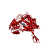
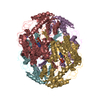
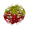
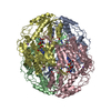
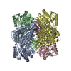
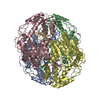
- PDB-3k2w: CRYSTAL STRUCTURE OF betaine-aldehyde dehydrogenase FROM Pseudoal... -
+
Open data
ID or keywords:
Loading...
-
Basic information
Entry
Database: PDB / ID: 3k2w
Title
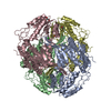
CRYSTAL STRUCTURE OF betaine-aldehyde dehydrogenase FROM Pseudoalteromonas atlantica T6c
Components
Betaine-aldehyde dehydrogenase
Keywords
OXIDOREDUCTASE / STRUCTURAL GENOMICS / PSI-2 / ALDEHYDE DEHYDROGENASE / PROTEIN STRUCTURE INITIATIVE / NEW YORK SGX RESEARCH CENTER FOR STRUCTURAL GENOMICS / NYSGXRC
Protocol: SINGLE WAVELENGTH / Monochromatic (M) / Laue (L): M / Scattering type: x-ray
Radiation wavelength
Wavelength: 0.9789 Å / Relative weight: 1
Reflection
Resolution: 1.9→40 Å / Num. obs: 310867 / % possible obs: 98.8 % / Observed criterion σ(I): -5 / Redundancy: 2.6 % / Biso Wilson estimate: 30.32 Å2 / Rsym value: 0.113 / Net I/σ(I): 3.7
Reflection shell
Resolution: 1.9→1.97 Å / Redundancy: 2.5 % / Mean I/σ(I) obs: 0.5 / % possible all: 97
-
Processing
Software
Name
Version
Classification
SHELX
modelbuilding
REFMAC
5.3.0034
refinement
HKL-2000
datareduction
HKL-2000
datascaling
SHELX
phasing
Refinement
Method to determine structure: SAD / Resolution: 1.9→20 Å / Cor.coef. Fo:Fc: 0.958 / Cor.coef. Fo:Fc free: 0.924 / SU B: 5.59 / SU ML: 0.159 / Cross valid method: THROUGHOUT / ESU R: 0.176 / ESU R Free: 0.174 / Stereochemistry target values: MAXIMUM LIKELIHOOD / Details: HYDROGENS HAVE BEEN ADDED IN THE RIDING POSITIONS
Rfactor
Num. reflection
% reflection
Selection details
Rfree
0.274
9231
3 %
RANDOM
Rwork
0.21
-
-
-
obs
0.212
293687
97.6 %
-
Solvent computation
Ion probe radii: 0.8 Å / Shrinkage radii: 0.8 Å / VDW probe radii: 1.2 Å / Solvent model: MASK
In the structure databanks used in Yorodumi, some data are registered as the other names, "COVID-19 virus" and "2019-nCoV". Here are the details of the virus and the list of structure data.
Jan 31, 2019. EMDB accession codes are about to change! (news from PDBe EMDB page)
EMDB accession codes are about to change! (news from PDBe EMDB page)
The allocation of 4 digits for EMDB accession codes will soon come to an end. Whilst these codes will remain in use, new EMDB accession codes will include an additional digit and will expand incrementally as the available range of codes is exhausted. The current 4-digit format prefixed with “EMD-” (i.e. EMD-XXXX) will advance to a 5-digit format (i.e. EMD-XXXXX), and so on. It is currently estimated that the 4-digit codes will be depleted around Spring 2019, at which point the 5-digit format will come into force.
The EM Navigator/Yorodumi systems omit the EMD- prefix.
Related info.:Q: What is EMD? / ID/Accession-code notation in Yorodumi/EM Navigator
Yorodumi is a browser for structure data from EMDB, PDB, SASBDB, etc.
This page is also the successor to EM Navigator detail page, and also detail information page/front-end page for Omokage search.
The word "yorodu" (or yorozu) is an old Japanese word meaning "ten thousand". "mi" (miru) is to see.
Related info.:EMDB / PDB / SASBDB / Comparison of 3 databanks / Yorodumi Search / Aug 31, 2016. New EM Navigator & Yorodumi / Yorodumi Papers / Jmol/JSmol / Function and homology information / Changes in new EM Navigator and Yorodumi
 Movie
Movie Controller

 Yorodumi
 Open data
 Basic information
 Components
 Keywords
 Function and homology information
 Pseudoalteromonas atlantica T6c (bacteria)
 X-RAY DIFFRACTION /
  Authors
 Citation
 Structure visualization
 Downloads & links
 Other downloads
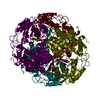


 PDBj
PDBj

 Assembly
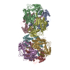


 Mass: 92.094 Da / Num. of mol.: 17 / Source method: obtained synthetically / Formula: C3H8O3
 Mass: 35.453 Da / Num. of mol.: 8 / Source method: obtained synthetically / Formula: Cl
 Mass: 59.044 Da / Num. of mol.: 6 / Source method: obtained synthetically / Formula: C2H3O2
 Mass: 18.015 Da / Num. of mol.: 1612 / Source method: isolated from a natural source / Formula: H2O
 Sample preparation
 / Beamline: X29A / Wavelength: 0.9789
  Processing