 Movie
Movie Controller

[English] 日本語
 Yorodumi
- PDB-5iqm: Crystal structure of the E. coli type 1 pilus subunit FimG (engin... -
+ Open data

- Basic information
| Entry | Database: PDB / ID: 5iqm | ||||||
|---|---|---|---|---|---|---|---|
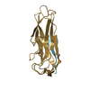
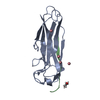
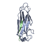
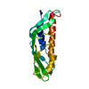
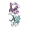
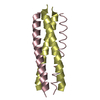
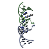
| Title | Crystal structure of the E. coli type 1 pilus subunit FimG (engineered variant with substitution Q134E; N-terminal FimG residues 1-12 truncated) in complex with the donor strand peptide DsF_T4R-T6R-D13N | ||||||
 Components Components |
| ||||||
 Keywords Keywords | CELL ADHESION / Complex / Protein / FimGt | ||||||
| Function / homology |  Function and homology information Function and homology informationpilus assembly / pilus tip / mechanosensory behavior / cell adhesion involved in single-species biofilm formation / pilus / cell-substrate adhesion / cell adhesion Similarity search - Function | ||||||
| Biological species |  | ||||||
| Method |  X-RAY DIFFRACTION / SYNCHROTRON / MOLECULAR REPLACEMENT / Resolution: 1.5 Å X-RAY DIFFRACTION / SYNCHROTRON / MOLECULAR REPLACEMENT / Resolution: 1.5 Å | ||||||
 Authors Authors | Giese, C. / Eras, J. / Kern, A. / Scharer, M.A. / Capitani, G. / Glockshuber, R. | ||||||
 Citation Citation | Journal: Angew.Chem.Int.Ed.Engl. / Year: 2016 Title: Accelerating the Association of the Most Stable Protein-Ligand Complex by More than Two Orders of Magnitude. Authors: Giese, C. / Eras, J. / Kern, A. / Scharer, M.A. / Capitani, G. / Glockshuber, R. | ||||||
| History |
|
- Structure visualization
| Structure viewer | Molecule: MolmilJmol/JSmol |
|---|
- Downloads & links
-Download
| PDBx/mmCIF format | 5iqm.cif.gz | 127.3 KB | Display | PDBx/mmCIF format |
|---|---|---|---|---|
| PDB format | pdb5iqm.ent.gz | 98.4 KB | Display | PDB format |
| PDBx/mmJSON format | 5iqm.json.gz | Tree view | PDBx/mmJSON format | |
| Others |  Other downloads Other downloads |
-Validation report
| Arichive directory | https://data.pdbj.org/pub/pdb/validation_reports/iq/5iqmftp://data.pdbj.org/pub/pdb/validation_reports/iq/5iqm | HTTPS FTP |
|---|
-Related structure data
| Related structure data |  5iqnC  5iqoC 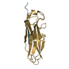 3bfqS S: Starting model for refinement C: citing same article ( |
|---|---|
| Similar structure data |

-Links
 PDBj
PDBj
- Assembly
| Deposited unit | 
| ||||||||
|---|---|---|---|---|---|---|---|---|---|
| 1 | 
| ||||||||
| 2 | 
| ||||||||
| Unit cell |
|
-Components
| #1: Protein | Mass: 13666.820 Da / Num. of mol.: 2 / Fragment: UNP residues 36-167 Source method: isolated from a genetically manipulated source Source: (gene. exp.) #2: Protein/peptide | Mass: 1750.963 Da / Num. of mol.: 2 / Fragment: UNP residues 23-37 / Source method: obtained synthetically / Source: (synth.) #3: Chemical | ChemComp-CO /   Mass: 58.933 Da / Num. of mol.: 4 / Source method: obtained synthetically / Formula: Co Mass: 58.933 Da / Num. of mol.: 4 / Source method: obtained synthetically / Formula: Co#4: Water | ChemComp-HOH / |  Mass: 18.015 Da / Num. of mol.: 419 / Source method: isolated from a natural source / Formula: H2O Mass: 18.015 Da / Num. of mol.: 419 / Source method: isolated from a natural source / Formula: H2OHas protein modification | Y | |
|---|
-Experimental details
-Experiment
| Experiment | Method: X-RAY DIFFRACTION / Number of used crystals: 1 |
|---|
- Sample preparation
| Crystal | Density Matthews: 2.32 Å3/Da / Density % sol: 47.09 % |
|---|---|
| Crystal grow | Temperature: 293 K / Method: vapor diffusion, sitting drop / Details: MOPS-NaOH pH 7.0 (RT), PEG-1500, NaCl, CoCl2 |
-Data collection
| Diffraction | Mean temperature: 100 K |
|---|---|
| Diffraction source | Source: SYNCHROTRON / Site: SLS  / Beamline: X06DA / Wavelength: 1 Å / Beamline: X06DA / Wavelength: 1 Å |
| Detector | Type: DECTRIS PILATUS 2M-F / Detector: PIXEL / Date: Jun 2, 2012 |
| Radiation | Protocol: SINGLE WAVELENGTH / Monochromatic (M) / Laue (L): M / Scattering type: x-ray |
| Radiation wavelength | Wavelength: 1 Å / Relative weight: 1 |
| Reflection | Resolution: 1.5→50 Å / Num. obs: 88780 / % possible obs: 99.6 % / Redundancy: 6.9 % / Rmerge(I) obs: 0.1 / Net I/σ(I): 14 |
| Reflection shell | Lowest resolution: 1.54 Å / Redundancy: 1.5 % / Rmerge(I) obs: 1.182 |
- Processing
| Software |
| ||||||||||||||||||||||||||||||||||||||||||||||||||||||||||||||||||||||||||||||||||||||||||||||||||
|---|---|---|---|---|---|---|---|---|---|---|---|---|---|---|---|---|---|---|---|---|---|---|---|---|---|---|---|---|---|---|---|---|---|---|---|---|---|---|---|---|---|---|---|---|---|---|---|---|---|---|---|---|---|---|---|---|---|---|---|---|---|---|---|---|---|---|---|---|---|---|---|---|---|---|---|---|---|---|---|---|---|---|---|---|---|---|---|---|---|---|---|---|---|---|---|---|---|---|---|
| Refinement | Method to determine structure: MOLECULAR REPLACEMENT Starting model: 3bfq Resolution: 1.5→47.889 Å / SU ML: 0.15 / Cross valid method: THROUGHOUT / σ(F): 1.37 / Phase error: 17.4
| ||||||||||||||||||||||||||||||||||||||||||||||||||||||||||||||||||||||||||||||||||||||||||||||||||
| Solvent computation | Shrinkage radii: 0.9 Å / VDW probe radii: 1.11 Å | ||||||||||||||||||||||||||||||||||||||||||||||||||||||||||||||||||||||||||||||||||||||||||||||||||
| Refinement step | Cycle: LAST / Resolution: 1.5→47.889 Å
| ||||||||||||||||||||||||||||||||||||||||||||||||||||||||||||||||||||||||||||||||||||||||||||||||||
| Refine LS restraints |
| ||||||||||||||||||||||||||||||||||||||||||||||||||||||||||||||||||||||||||||||||||||||||||||||||||
| LS refinement shell |
|
