 Movie
Movie Controller

[English] 日本語
 Yorodumi
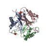
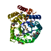
- PDB-5ioh: RepoMan-PP1a (protein phosphatase 1, alpha isoform) holoenzyme complex -
+ Open data

- Basic information
| Entry | Database: PDB / ID: 5ioh | |||||||||
|---|---|---|---|---|---|---|---|---|---|---|
| Title | RepoMan-PP1a (protein phosphatase 1, alpha isoform) holoenzyme complex | |||||||||
 Components Components |
| |||||||||
 Keywords Keywords | HYDROLASE/PROTEIN BINDING / PP1 alpha / RepoMan / Ki-67 / Phosphatase / HYDROLASE-PROTEIN BINDING complex | |||||||||
| Function / homology |  Function and homology information Function and homology informationregulation of glycogen catabolic process / positive regulation of termination of RNA polymerase II transcription, poly(A)-coupled / PTW/PP1 phosphatase complex / protein phosphatase type 1 complex / glycogen granule / RNA polymerase II promoter clearance / RNA polymerase II CTD heptapeptide repeat S5 phosphatase activity / cadherin binding involved in cell-cell adhesion / protein phosphatase 1 binding / regulation of translational initiation in response to stress ...regulation of glycogen catabolic process / positive regulation of termination of RNA polymerase II transcription, poly(A)-coupled / PTW/PP1 phosphatase complex / protein phosphatase type 1 complex / glycogen granule / RNA polymerase II promoter clearance / RNA polymerase II CTD heptapeptide repeat S5 phosphatase activity / cadherin binding involved in cell-cell adhesion / protein phosphatase 1 binding / regulation of translational initiation in response to stress / protein phosphatase regulator activity / positive regulation of extrinsic apoptotic signaling pathway in absence of ligand / regulation of canonical Wnt signaling pathway / protein dephosphorylation / Phosphorylation and nuclear translocation of the CRY:PER:kinase complex / glycogen metabolic process / regulation of mitotic nuclear division / Triglyceride catabolism / branching morphogenesis of an epithelial tube / protein-serine/threonine phosphatase / entrainment of circadian clock by photoperiod / protein serine/threonine phosphatase activity / telomere maintenance in response to DNA damage / phosphatase activity / Maturation of hRSV A proteins / negative regulation of transcription elongation by RNA polymerase II / transition metal ion binding / positive regulation of glycogen biosynthetic process / DARPP-32 events / ribonucleoprotein complex binding / phosphoprotein phosphatase activity / lung development / Downregulation of TGF-beta receptor signaling / circadian regulation of gene expression / adherens junction / positive regulation of transcription elongation by RNA polymerase II / centriole / sperm end piece / chromosome segregation / regulation of circadian rhythm / response to lead ion / chromosome / presynapse / sperm midpiece / dendritic spine / perikaryon / protein stabilization / iron ion binding / cell division / nucleolus / glutamatergic synapse / endoplasmic reticulum / extracellular exosome / nucleoplasm / nucleus / plasma membrane / cytoplasm / cytosol Similarity search - Function | |||||||||
| Biological species |  Homo sapiens (human) Homo sapiens (human) | |||||||||
| Method |  X-RAY DIFFRACTION / SYNCHROTRON / MOLECULAR REPLACEMENT / Resolution: 2.566 Å X-RAY DIFFRACTION / SYNCHROTRON / MOLECULAR REPLACEMENT / Resolution: 2.566 Å | |||||||||
 Authors Authors | Kumar, G.S. / Peti, W. / Page, R. | |||||||||
| Funding support |  United States, 2items United States, 2items
| |||||||||
 Citation Citation | Journal: Elife / Year: 2016 Title: The Ki-67 and RepoMan mitotic phosphatases assemble via an identical, yet novel mechanism. Authors: Kumar, G.S. / Gokhan, E. / De Munter, S. / Bollen, M. / Vagnarelli, P. / Peti, W. / Page, R. | |||||||||
| History |
|
- Structure visualization
| Structure viewer | Molecule: MolmilJmol/JSmol |
|---|
- Downloads & links
-Download
| PDBx/mmCIF format | 5ioh.cif.gz | 140.8 KB | Display | PDBx/mmCIF format |
|---|---|---|---|---|
| PDB format | pdb5ioh.ent.gz | 109.1 KB | Display | PDB format |
| PDBx/mmJSON format | 5ioh.json.gz | Tree view | PDBx/mmJSON format | |
| Others |  Other downloads Other downloads |
-Validation report
| Arichive directory | https://data.pdbj.org/pub/pdb/validation_reports/io/5iohftp://data.pdbj.org/pub/pdb/validation_reports/io/5ioh | HTTPS FTP |
|---|
-Related structure data
| Related structure data |  5inbC  5j28C  4movS C: citing same article ( S: Starting model for refinement |
|---|---|
| Similar structure data | |
| Other databases |
|


-Links
 PDBj
PDBj


- Assembly
| Deposited unit | 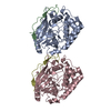
| ||||||||
|---|---|---|---|---|---|---|---|---|---|
| 1 | 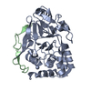
| ||||||||
| 2 | 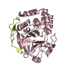
| ||||||||
| Unit cell |
|
-Components
| #1: Protein | Mass: 34162.148 Da / Num. of mol.: 2 / Fragment: UNP residues 7-300 Source method: isolated from a genetically manipulated source Source: (gene. exp.) Homo sapiens (human) / Gene: PPP1CA, PPP1A / Plasmid: RP1B / Production host:  References: UniProt: P62136, protein-serine/threonine phosphatase #2: Protein | Mass: 7023.053 Da / Num. of mol.: 2 / Fragment: UNP residues 383-441 Source method: isolated from a genetically manipulated source Source: (gene. exp.) Homo sapiens (human) / Gene: CDCA2 / Production host: #3: Water | ChemComp-HOH / |  Mass: 18.015 Da / Num. of mol.: 152 / Source method: isolated from a natural source / Formula: H2O Mass: 18.015 Da / Num. of mol.: 152 / Source method: isolated from a natural source / Formula: H2O |
|---|
-Experimental details
-Experiment
| Experiment | Method: X-RAY DIFFRACTION / Number of used crystals: 1 |
|---|
- Sample preparation
| Crystal | Density Matthews: 2.05 Å3/Da / Density % sol: 40.04 % |
|---|---|
| Crystal grow | Temperature: 277 K / Method: vapor diffusion, sitting drop / pH: 4 / Details: 100 mM Sodium Malonate, 12% PEG 3350 |
-Data collection
| Diffraction | Mean temperature: 100 K |
|---|---|
| Diffraction source | Source: SYNCHROTRON / Site: NSLS / Beamline: X25 / Wavelength: 1.1 Å |
| Detector | Type: DECTRIS PILATUS 6M / Detector: PIXEL / Date: Nov 12, 2013 |
| Radiation | Monochromator: Si(111) / Protocol: SINGLE WAVELENGTH / Monochromatic (M) / Laue (L): M / Scattering type: x-ray |
| Radiation wavelength | Wavelength: 1.1 Å / Relative weight: 1 |
| Reflection | Resolution: 2.566→50 Å / Num. obs: 22441 / % possible obs: 99.3 % / Redundancy: 11.5 % / Rmerge(I) obs: 0.017 / Net I/σ(I): 15.3 |
| Reflection shell | Resolution: 2.6→2.64 Å / Redundancy: 5.9 % / Mean I/σ(I) obs: 2.4 / % possible all: 90.4 |
- Processing
| Software |
| |||||||||||||||||||||||||||||||||||||||||||||||||||||||||||||||
|---|---|---|---|---|---|---|---|---|---|---|---|---|---|---|---|---|---|---|---|---|---|---|---|---|---|---|---|---|---|---|---|---|---|---|---|---|---|---|---|---|---|---|---|---|---|---|---|---|---|---|---|---|---|---|---|---|---|---|---|---|---|---|---|---|
| Refinement | Method to determine structure: MOLECULAR REPLACEMENT Starting model: 4MOV Resolution: 2.566→46.545 Å / SU ML: 0.31 / Cross valid method: THROUGHOUT / σ(F): 1.36 / Phase error: 21.16 / Stereochemistry target values: ML
| |||||||||||||||||||||||||||||||||||||||||||||||||||||||||||||||
| Solvent computation | Shrinkage radii: 0.9 Å / VDW probe radii: 1.11 Å / Solvent model: FLAT BULK SOLVENT MODEL | |||||||||||||||||||||||||||||||||||||||||||||||||||||||||||||||
| Refinement step | Cycle: LAST / Resolution: 2.566→46.545 Å
| |||||||||||||||||||||||||||||||||||||||||||||||||||||||||||||||
| Refine LS restraints |
| |||||||||||||||||||||||||||||||||||||||||||||||||||||||||||||||
| LS refinement shell |
|
