 Movie
Movie Controller

[English] 日本語
 Yorodumi
- PDB-5j28: Ki67-PP1g (protein phosphatase 1, gamma isoform) holoenzyme complex -
+ Open data

- Basic information
| Entry | Database: PDB / ID: 5j28 | |||||||||
|---|---|---|---|---|---|---|---|---|---|---|
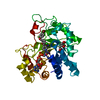
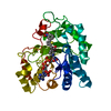
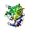
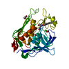
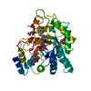
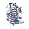
| Title | Ki67-PP1g (protein phosphatase 1, gamma isoform) holoenzyme complex | |||||||||
 Components Components |
| |||||||||
 Keywords Keywords | HYDROLASE/PROTEIN BINDING / PP1 gamma / RepoMan / Ki-67 / Phosphatase / HYDROLASE-PROTEIN BINDING complex | |||||||||
| Function / homology |  Function and homology information Function and homology informationPTW/PP1 phosphatase complex / regulation of nucleocytoplasmic transport / regulation of chromatin organization / protein phosphatase 1 binding / lamin binding / SHOC2 M1731 mutant abolishes MRAS complex function / Gain-of-function MRAS complexes activate RAF signaling / protein dephosphorylation / Phosphorylation and nuclear translocation of the CRY:PER:kinase complex / glycogen metabolic process ...PTW/PP1 phosphatase complex / regulation of nucleocytoplasmic transport / regulation of chromatin organization / protein phosphatase 1 binding / lamin binding / SHOC2 M1731 mutant abolishes MRAS complex function / Gain-of-function MRAS complexes activate RAF signaling / protein dephosphorylation / Phosphorylation and nuclear translocation of the CRY:PER:kinase complex / glycogen metabolic process / regulation of mitotic nuclear division / Triglyceride catabolism / protein-serine/threonine phosphatase / entrainment of circadian clock by photoperiod / protein serine/threonine phosphatase activity / cleavage furrow / phosphatase activity / Maturation of hRSV A proteins / microtubule organizing center / mitotic sister chromatid segregation / positive regulation of glial cell proliferation / blastocyst development / condensed chromosome / phosphoprotein phosphatase activity / Amplification of signal from unattached kinetochores via a MAD2 inhibitory signal / Mitotic Prometaphase / EML4 and NUDC in mitotic spindle formation / Resolution of Sister Chromatid Cohesion / Downregulation of TGF-beta receptor signaling / circadian regulation of gene expression / molecular condensate scaffold activity / chromosome segregation / RAF activation / RHO GTPases Activate Formins / regulation of circadian rhythm / kinetochore / neuron differentiation / Separation of Sister Chromatids / MAPK cascade / chromosome / presynapse / midbody / spermatogenesis / dendritic spine / mitochondrial outer membrane / cell population proliferation / nuclear speck / protein domain specific binding / cell division / focal adhesion / protein kinase binding / protein-containing complex binding / nucleolus / glutamatergic synapse / protein-containing complex / mitochondrion / DNA binding / RNA binding / nucleoplasm / ATP binding / membrane / metal ion binding / nucleus / cytoplasm / cytosol Similarity search - Function | |||||||||
| Biological species |  Homo sapiens (human) Homo sapiens (human) | |||||||||
| Method |  X-RAY DIFFRACTION / SYNCHROTRON / MOLECULAR REPLACEMENT / Resolution: 2 Å X-RAY DIFFRACTION / SYNCHROTRON / MOLECULAR REPLACEMENT / Resolution: 2 Å | |||||||||
 Authors Authors | Kumar, G.S. / Peti, W. / Page, R. | |||||||||
| Funding support |  United States, 2items United States, 2items
| |||||||||
 Citation Citation | Journal: Elife / Year: 2016 Title: The Ki-67 and RepoMan mitotic phosphatases assemble via an identical, yet novel mechanism. Authors: Kumar, G.S. / Gokhan, E. / De Munter, S. / Bollen, M. / Vagnarelli, P. / Peti, W. / Page, R. | |||||||||
| History |
|
- Structure visualization
| Structure viewer | Molecule: MolmilJmol/JSmol |
|---|
- Downloads & links
-Download
| PDBx/mmCIF format | 5j28.cif.gz | 146.4 KB | Display | PDBx/mmCIF format |
|---|---|---|---|---|
| PDB format | pdb5j28.ent.gz | 112.7 KB | Display | PDB format |
| PDBx/mmJSON format | 5j28.json.gz | Tree view | PDBx/mmJSON format | |
| Others |  Other downloads Other downloads |
-Validation report
| Arichive directory | https://data.pdbj.org/pub/pdb/validation_reports/j2/5j28ftp://data.pdbj.org/pub/pdb/validation_reports/j2/5j28 | HTTPS FTP |
|---|
-Related structure data
| Related structure data | 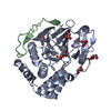 5inbSC  5iohC S: Starting model for refinement C: citing same article ( |
|---|---|
| Similar structure data | |
| Other databases |
|


-Links
 PDBj
PDBj


- Assembly
| Deposited unit | 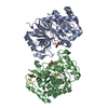
| ||||||||
|---|---|---|---|---|---|---|---|---|---|
| 1 | 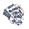
| ||||||||
| 2 | 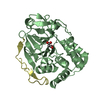
| ||||||||
| Unit cell |
|
-Components
| #1: Protein | Mass: 35044.363 Da / Num. of mol.: 2 / Fragment: UNP residues 7-308 Source method: isolated from a genetically manipulated source Source: (gene. exp.) Homo sapiens (human) / Gene: PPP1CC / Plasmid: RP1B / Production host:  References: UniProt: P36873, protein-serine/threonine phosphatase #2: Protein/peptide | Mass: 5187.017 Da / Num. of mol.: 2 / Fragment: UNP residues 496-536 / Mutation: T525M Source method: isolated from a genetically manipulated source Source: (gene. exp.) Homo sapiens (human) / Gene: MKI67 / Plasmid: pet-M30 MBP / Production host: #3: Chemical |   Mass: 102.046 Da / Num. of mol.: 2 / Source method: obtained synthetically / Formula: C3H2O4 Mass: 102.046 Da / Num. of mol.: 2 / Source method: obtained synthetically / Formula: C3H2O4#4: Chemical | ChemComp-NA / |   Mass: 22.990 Da / Num. of mol.: 1 / Source method: obtained synthetically / Formula: Na Mass: 22.990 Da / Num. of mol.: 1 / Source method: obtained synthetically / Formula: Na#5: Water | ChemComp-HOH / |  Mass: 18.015 Da / Num. of mol.: 232 / Source method: isolated from a natural source / Formula: H2O Mass: 18.015 Da / Num. of mol.: 232 / Source method: isolated from a natural source / Formula: H2O |
|---|
-Experimental details
-Experiment
| Experiment | Method: X-RAY DIFFRACTION / Number of used crystals: 1 |
|---|
- Sample preparation
| Crystal | Density Matthews: 3.06 Å3/Da / Density % sol: 59.8 % |
|---|---|
| Crystal grow | Temperature: 277 K / Method: vapor diffusion, hanging drop / pH: 4 / Details: 1.9 M Sodium Malonate pH 4.0 |
-Data collection
| Diffraction | Mean temperature: 100 K |
|---|---|
| Diffraction source | Source: SYNCHROTRON / Site: SSRL / Beamline: BL12-2 / Wavelength: 0.9795 Å |
| Detector | Type: DECTRIS PILATUS 6M / Detector: PIXEL / Date: Mar 19, 2016 |
| Radiation | Monochromator: Liquid nitrogen-cooled double crystal, non fixed exit slit Protocol: SINGLE WAVELENGTH / Monochromatic (M) / Laue (L): M / Scattering type: x-ray |
| Radiation wavelength | Wavelength: 0.9795 Å / Relative weight: 1 |
| Reflection | Resolution: 2→39.331 Å / Num. obs: 64993 / % possible obs: 100 % / Observed criterion σ(I): 3.1 / Redundancy: 10.4 % / Rmerge(I) obs: 0.092 / Net I/σ(I): 20.5 |
| Reflection shell | Resolution: 2→2.05 Å / Redundancy: 10.3 % / Rmerge(I) obs: 0.0861 / Mean I/σ(I) obs: 3.1 / % possible all: 92.2 |
- Processing
| Software |
| |||||||||||||||||||||||||||||||||||||||||||||||||||||||||||||||||||||||||||||||||||||||||||||||||||||||||
|---|---|---|---|---|---|---|---|---|---|---|---|---|---|---|---|---|---|---|---|---|---|---|---|---|---|---|---|---|---|---|---|---|---|---|---|---|---|---|---|---|---|---|---|---|---|---|---|---|---|---|---|---|---|---|---|---|---|---|---|---|---|---|---|---|---|---|---|---|---|---|---|---|---|---|---|---|---|---|---|---|---|---|---|---|---|---|---|---|---|---|---|---|---|---|---|---|---|---|---|---|---|---|---|---|---|---|
| Refinement | Method to determine structure: MOLECULAR REPLACEMENT Starting model: 5INB Resolution: 2→39.331 Å / Cross valid method: THROUGHOUT / σ(F): 1.35 / Phase error: 20.23 / Stereochemistry target values: TWIN_LSQ_F
| |||||||||||||||||||||||||||||||||||||||||||||||||||||||||||||||||||||||||||||||||||||||||||||||||||||||||
| Solvent computation | Shrinkage radii: 0.9 Å / VDW probe radii: 1.11 Å / Solvent model: FLAT BULK SOLVENT MODEL | |||||||||||||||||||||||||||||||||||||||||||||||||||||||||||||||||||||||||||||||||||||||||||||||||||||||||
| Refinement step | Cycle: LAST / Resolution: 2→39.331 Å
| |||||||||||||||||||||||||||||||||||||||||||||||||||||||||||||||||||||||||||||||||||||||||||||||||||||||||
| Refine LS restraints |
| |||||||||||||||||||||||||||||||||||||||||||||||||||||||||||||||||||||||||||||||||||||||||||||||||||||||||
| LS refinement shell |
|
