 Movie
Movie Controller

[English] 日本語
 Yorodumi
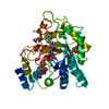
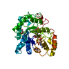
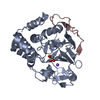
- PDB-1nar: CRYSTAL STRUCTURE OF NARBONIN REFINED AT 1.8 ANGSTROMS RESOLUTION -
+ Open data

- Basic information
| Entry | Database: PDB / ID: 1nar | ||||||
|---|---|---|---|---|---|---|---|
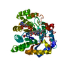
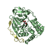
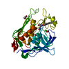
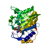
| Title | CRYSTAL STRUCTURE OF NARBONIN REFINED AT 1.8 ANGSTROMS RESOLUTION | ||||||
 Components Components | NARBONIN | ||||||
 Keywords Keywords | PLANT SEED PROTEIN | ||||||
| Function / homology |  Function and homology information Function and homology information | ||||||
| Biological species |  Vicia narbonensis (plant) Vicia narbonensis (plant) | ||||||
| Method |  X-RAY DIFFRACTION / Resolution: 1.8 Å X-RAY DIFFRACTION / Resolution: 1.8 Å | ||||||
 Authors Authors | Hennig, M. / Schlesier, B. / Wilson, K.S. | ||||||
 Citation Citation | Journal: Acta Crystallogr.,Sect.D / Year: 1995 Title: Crystal structure of narbonin at 1.8 A resolution. Authors: Hennig, M. / Pfeffer-Hennig, S. / Dauter, Z. / Wilson, K.S. / Schlesier, B. / Nong, V.H. #1: Journal: FEBS Lett. / Year: 1992Title: A Tim Barrel Protein without Enzymatic Activity? Crystal Structure of Narbonin at 1.8 Angstroms Resolution Authors: Hennig, M. / Schlesier, B. / Dauter, Z. / Pfeffer, S. / Betzel, C. / Hoehne, W.E. / Wilson, K.S. #2: Journal: J.Mol.Biol. / Year: 1990Title: Narbonin, a 2S Globulin from Vicia Narbonensis L. Crystallization and Preliminary Crystallographic Data Authors: Hennig, M. / Schlesier, B. / Pfeffer, S. / Hoehne, W.E. | ||||||
| History |
| ||||||
| Remark 700 | SHEET THE SHEET PRESENTED AS *A* ON SHEET RECORDS BELOW IS ACTUALLY AN EIGHT-STRANDED BETA-BARREL. ...SHEET THE SHEET PRESENTED AS *A* ON SHEET RECORDS BELOW IS ACTUALLY AN EIGHT-STRANDED BETA-BARREL. THIS IS REPRESENTED BY A NINE-STRANDED SHEET IN WHICH THE FIRST AND LAST STRANDS ARE IDENTICAL. |
- Structure visualization
| Structure viewer | Molecule: MolmilJmol/JSmol |
|---|
- Downloads & links
-Download
| PDBx/mmCIF format | 1nar.cif.gz | 82.4 KB | Display | PDBx/mmCIF format |
|---|---|---|---|---|
| PDB format | pdb1nar.ent.gz | 61.2 KB | Display | PDB format |
| PDBx/mmJSON format | 1nar.json.gz | Tree view | PDBx/mmJSON format | |
| Others |  Other downloads Other downloads |
-Validation report
| Arichive directory | https://data.pdbj.org/pub/pdb/validation_reports/na/1narftp://data.pdbj.org/pub/pdb/validation_reports/na/1nar | HTTPS FTP |
|---|
-Related structure data
| Similar structure data |
|---|


-Links
 PDBj
PDBj- Assembly
| Deposited unit | 
| ||||||||
|---|---|---|---|---|---|---|---|---|---|
| 1 |
| ||||||||
| Unit cell |
| ||||||||
| Atom site foot note | 1: GLY 38 - PHE 39 OMEGA = 9.34 PEPTIDE BOND DEVIATES SIGNIFICANTLY FROM TRANS CONFORMATION 2: CIS PROLINE - PRO 139 3: TRP 261 - ASN 262 OMEGA = 12.67 PEPTIDE BOND DEVIATES SIGNIFICANTLY FROM TRANS CONFORMATION |
-Components
| #1: Protein | Mass: 33140.184 Da / Num. of mol.: 1 Source method: isolated from a genetically manipulated source Source: (gene. exp.) Vicia narbonensis (plant) / References: UniProt: Q08884 |
|---|---|
| #2: Water | ChemComp-HOH /  Mass: 18.015 Da / Num. of mol.: 513 / Source method: isolated from a natural source / Formula: H2O Mass: 18.015 Da / Num. of mol.: 513 / Source method: isolated from a natural source / Formula: H2O |
| Sequence details | THE AMINO ACID SEQUENCE WAS DETERMINED BY INTERPRETATION OF THE ELECTRON DENSITY (REF. 1). FOR ...THE AMINO ACID SEQUENCE WAS DETERMINED |
-Experimental details
-Experiment
| Experiment | Method: X-RAY DIFFRACTION |
|---|
- Sample preparation
| Crystal | Density Matthews: 2.34 Å3/Da / Density % sol: 47.48 % | ||||||||||||||||||||||||||||||||||||||||||
|---|---|---|---|---|---|---|---|---|---|---|---|---|---|---|---|---|---|---|---|---|---|---|---|---|---|---|---|---|---|---|---|---|---|---|---|---|---|---|---|---|---|---|---|
| Crystal grow | *PLUS pH: 9.5 / Method: vapor diffusion, sitting drop | ||||||||||||||||||||||||||||||||||||||||||
| Components of the solutions | *PLUS
|
-Data collection
| Radiation | Scattering type: x-ray |
|---|---|
| Radiation wavelength | Relative weight: 1 |
| Reflection | *PLUS Highest resolution: 1.8 Å / Lowest resolution: 10 Å / Num. obs: 27227 / % possible obs: 95.5 % / Num. measured all: 124224 / Rmerge(I) obs: 0.056 |
| Reflection shell | *PLUS Highest resolution: 1.8 Å / Lowest resolution: 1.85 Å / Rmerge(I) obs: 0.078 |
- Processing
| Software | Name: PROLSQ / Classification: refinement | ||||||||||||||||||||||||||||||||||||||||||||||||||||||||||||||||||||||||||||||||||||
|---|---|---|---|---|---|---|---|---|---|---|---|---|---|---|---|---|---|---|---|---|---|---|---|---|---|---|---|---|---|---|---|---|---|---|---|---|---|---|---|---|---|---|---|---|---|---|---|---|---|---|---|---|---|---|---|---|---|---|---|---|---|---|---|---|---|---|---|---|---|---|---|---|---|---|---|---|---|---|---|---|---|---|---|---|---|
| Refinement | Resolution: 1.8→10 Å / σ(F): 0 /
| ||||||||||||||||||||||||||||||||||||||||||||||||||||||||||||||||||||||||||||||||||||
| Refinement step | Cycle: LAST / Resolution: 1.8→10 Å
| ||||||||||||||||||||||||||||||||||||||||||||||||||||||||||||||||||||||||||||||||||||
| Refine LS restraints |
| ||||||||||||||||||||||||||||||||||||||||||||||||||||||||||||||||||||||||||||||||||||
| Software | *PLUS Name: PROLSQ / Classification: refinement | ||||||||||||||||||||||||||||||||||||||||||||||||||||||||||||||||||||||||||||||||||||
| Refinement | *PLUS Highest resolution: 1.8 Å / Lowest resolution: 10 Å / Num. reflection all: 27227 / σ(F): 0 / Rfactor all: 0.159 | ||||||||||||||||||||||||||||||||||||||||||||||||||||||||||||||||||||||||||||||||||||
| Solvent computation | *PLUS | ||||||||||||||||||||||||||||||||||||||||||||||||||||||||||||||||||||||||||||||||||||
| Displacement parameters | *PLUS Biso mean: 21 Å2 |
