 Movie
Movie Controller

[English] 日本語
 Yorodumi
- PDB-5il0: Crystal structural of the METTL3-METTL14 complex for N6-adenosine... -
+ Open data

- Basic information
| Entry | Database: PDB / ID: 5il0 | ||||||
|---|---|---|---|---|---|---|---|
| Title | Crystal structural of the METTL3-METTL14 complex for N6-adenosine methylation | ||||||
 Components Components |
| ||||||
 Keywords Keywords | RNA BINDING PROTEIN / methyltransferase | ||||||
| Function / homology |  Function and homology information Function and homology informationnegative regulation of hematopoietic progenitor cell differentiation / mRNA m(6)A methyltransferase activity / positive regulation of cap-independent translational initiation / mRNA m6A methyltransferase / RNA N6-methyladenosine methyltransferase complex / RNA methylation / endothelial to hematopoietic transition / RNA methyltransferase activity / regulation of meiotic cell cycle / primary miRNA processing ...negative regulation of hematopoietic progenitor cell differentiation / mRNA m(6)A methyltransferase activity / positive regulation of cap-independent translational initiation / mRNA m6A methyltransferase / RNA N6-methyladenosine methyltransferase complex / RNA methylation / endothelial to hematopoietic transition / RNA methyltransferase activity / regulation of meiotic cell cycle / primary miRNA processing / forebrain radial glial cell differentiation / dosage compensation by inactivation of X chromosome / S-adenosyl-L-methionine binding / gliogenesis / regulation of hematopoietic stem cell differentiation / regulation of T cell differentiation / regulation of neuron differentiation / negative regulation of type I interferon-mediated signaling pathway / mRNA stabilization / oogenesis / mRNA modification / stem cell population maintenance / mRNA destabilization / Processing of Capped Intron-Containing Pre-mRNA / negative regulation of Notch signaling pathway / positive regulation of translation / mRNA splicing, via spliceosome / circadian rhythm / response to nutrient levels / mRNA processing / cellular response to UV / spermatogenesis / nuclear speck / nuclear body / protein heterodimerization activity / innate immune response / mRNA binding / DNA damage response / Golgi apparatus / nucleoplasm / nucleus / cytosol Similarity search - Function | ||||||
| Biological species |  Homo sapiens (human) Homo sapiens (human) | ||||||
| Method |  X-RAY DIFFRACTION / SYNCHROTRON / SAD / Resolution: 1.882 Å X-RAY DIFFRACTION / SYNCHROTRON / SAD / Resolution: 1.882 Å | ||||||
 Authors Authors | Wang, X. / Guan, Z. / Zou, T. / Yin, P. | ||||||
 Citation Citation | Journal: Nature / Year: 2016 Title: Structural basis of N6-adenosine methylation by the METTL3-METTL14 complex Authors: Wang, X. / Feng, J. / Xue, Y. / Guan, Z. / Zhang, D. / Liu, Z. / Gong, Z. / Wang, Q. / Huang, J. / Tang, C. / Zou, T. / Yin, P. | ||||||
| History |
|
- Structure visualization
| Structure viewer | Molecule: MolmilJmol/JSmol |
|---|
- Downloads & links
-Download
| PDBx/mmCIF format | 5il0.cif.gz | 231.1 KB | Display | PDBx/mmCIF format |
|---|---|---|---|---|
| PDB format | pdb5il0.ent.gz | 184.4 KB | Display | PDB format |
| PDBx/mmJSON format | 5il0.json.gz | Tree view | PDBx/mmJSON format | |
| Others |  Other downloads Other downloads |
-Validation report
| Arichive directory | https://data.pdbj.org/pub/pdb/validation_reports/il/5il0ftp://data.pdbj.org/pub/pdb/validation_reports/il/5il0 | HTTPS FTP |
|---|
-Related structure data


-Links
 PDBj
PDBj

- Assembly
| Deposited unit | 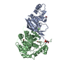
| ||||||||
|---|---|---|---|---|---|---|---|---|---|
| 1 |
| ||||||||
| Unit cell |
|
-Components
| #1: Protein | Mass: 24427.109 Da / Num. of mol.: 1 / Fragment: UNP residues 369-580 Source method: isolated from a genetically manipulated source Source: (gene. exp.) Homo sapiens (human) / Gene: METTL3 / Production host:  References: UniProt: Q86U44, mRNA (2'-O-methyladenosine-N6-)-methyltransferase | ||||||
|---|---|---|---|---|---|---|---|
| #2: Protein | Mass: 34794.402 Da / Num. of mol.: 1 / Fragment: UNP residues 109-408 Source method: isolated from a genetically manipulated source Source: (gene. exp.) Homo sapiens (human) / Gene: METTL14 / Production host: References: UniProt: Q9HCE5, mRNA (2'-O-methyladenosine-N6-)-methyltransferase | ||||||
| #3: Chemical |   Mass: 62.068 Da / Num. of mol.: 2 Mass: 62.068 Da / Num. of mol.: 2Source method: isolated from a genetically manipulated source Formula: C2H6O2 #4: Chemical | ChemComp-BR / |   Mass: 79.904 Da / Num. of mol.: 1 Mass: 79.904 Da / Num. of mol.: 1Source method: isolated from a genetically manipulated source Formula: Br #5: Water | ChemComp-HOH / |  Mass: 18.015 Da / Num. of mol.: 331 / Source method: isolated from a natural source / Formula: H2O Mass: 18.015 Da / Num. of mol.: 331 / Source method: isolated from a natural source / Formula: H2OHas protein modification | Y | |
-Experimental details
-Experiment
| Experiment | Method: X-RAY DIFFRACTION / Number of used crystals: 1 |
|---|
- Sample preparation
| Crystal | Density Matthews: 2.55 Å3/Da / Density % sol: 51.69 % |
|---|---|
| Crystal grow | Temperature: 291 K / Method: vapor diffusion, hanging drop / pH: 5.7 / Details: PEG 8000, sodium citrate |
-Data collection
| Diffraction | Mean temperature: 100 K |
|---|---|
| Diffraction source | Source: SYNCHROTRON / Site: SSRF  / Beamline: BL17U / Wavelength: 0.92 Å / Beamline: BL17U / Wavelength: 0.92 Å |
| Detector | Type: ADSC QUANTUM 315r / Detector: CCD / Date: Sep 8, 2015 |
| Radiation | Protocol: SINGLE WAVELENGTH / Monochromatic (M) / Laue (L): M / Scattering type: x-ray |
| Radiation wavelength | Wavelength: 0.92 Å / Relative weight: 1 |
| Reflection | Resolution: 1.88→50 Å / Num. obs: 50152 / % possible obs: 99.9 % / Redundancy: 14.6 % / Net I/σ(I): 37.5 |
- Processing
| Software |
| |||||||||||||||||||||||||||||||||||||||||||||||||||||||||||||||||||||||||||||||||||||||||||||||||||||||||||||||||||||||||||||||||||||
|---|---|---|---|---|---|---|---|---|---|---|---|---|---|---|---|---|---|---|---|---|---|---|---|---|---|---|---|---|---|---|---|---|---|---|---|---|---|---|---|---|---|---|---|---|---|---|---|---|---|---|---|---|---|---|---|---|---|---|---|---|---|---|---|---|---|---|---|---|---|---|---|---|---|---|---|---|---|---|---|---|---|---|---|---|---|---|---|---|---|---|---|---|---|---|---|---|---|---|---|---|---|---|---|---|---|---|---|---|---|---|---|---|---|---|---|---|---|---|---|---|---|---|---|---|---|---|---|---|---|---|---|---|---|---|
| Refinement | Method to determine structure: SAD / Resolution: 1.882→36.272 Å / SU ML: 0.18 / Cross valid method: FREE R-VALUE / σ(F): 1.34 / Phase error: 20.52
| |||||||||||||||||||||||||||||||||||||||||||||||||||||||||||||||||||||||||||||||||||||||||||||||||||||||||||||||||||||||||||||||||||||
| Solvent computation | Shrinkage radii: 0.9 Å / VDW probe radii: 1.11 Å | |||||||||||||||||||||||||||||||||||||||||||||||||||||||||||||||||||||||||||||||||||||||||||||||||||||||||||||||||||||||||||||||||||||
| Refinement step | Cycle: LAST / Resolution: 1.882→36.272 Å
| |||||||||||||||||||||||||||||||||||||||||||||||||||||||||||||||||||||||||||||||||||||||||||||||||||||||||||||||||||||||||||||||||||||
| Refine LS restraints |
| |||||||||||||||||||||||||||||||||||||||||||||||||||||||||||||||||||||||||||||||||||||||||||||||||||||||||||||||||||||||||||||||||||||
| LS refinement shell |
| |||||||||||||||||||||||||||||||||||||||||||||||||||||||||||||||||||||||||||||||||||||||||||||||||||||||||||||||||||||||||||||||||||||
| Refinement TLS params. | Method: refined / Origin x: 9.5129 Å / Origin y: 21.5197 Å / Origin z: 133.6862 Å
| |||||||||||||||||||||||||||||||||||||||||||||||||||||||||||||||||||||||||||||||||||||||||||||||||||||||||||||||||||||||||||||||||||||
| Refinement TLS group | Selection details: all |
