 Movie
Movie Controller

[English] 日本語
 Yorodumi
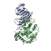
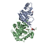
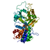
- PDB-1np7: Crystal Structure Analysis of Synechocystis sp. PCC6803 cryptochrome -
+ Open data

- Basic information
| Entry | Database: PDB / ID: 1np7 | ||||||
|---|---|---|---|---|---|---|---|
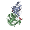
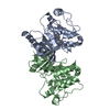
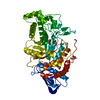
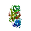
| Title | Crystal Structure Analysis of Synechocystis sp. PCC6803 cryptochrome | ||||||
 Components Components | DNA photolyase | ||||||
 Keywords Keywords | LYASE / protein with FAD cofactor | ||||||
| Function / homology |  Function and homology information Function and homology informationDNA photolyase activity / photoreactive repair / FAD binding / DNA binding Similarity search - Function | ||||||
| Biological species |  | ||||||
| Method |  X-RAY DIFFRACTION / SYNCHROTRON / MOLECULAR REPLACEMENT / Resolution: 1.9 Å X-RAY DIFFRACTION / SYNCHROTRON / MOLECULAR REPLACEMENT / Resolution: 1.9 Å | ||||||
 Authors Authors | Brudler, R. / Hitomi, K. / Daiyasu, H. / Toh, H. / Kucho, K. / Ishiura, M. / Kanehisa, M. / Roberts, V.A. / Todo, T. / Tainer, J.A. / Getzoff, E.D. | ||||||
 Citation Citation | Journal: Mol.Cell / Year: 2003 Title: Identification of a new cryptochrome class: structure, function, and evolution Authors: Brudler, R. / Hitomi, K. / Daiyasu, H. / Toh, H. / Kucho, K. / Ishiura, M. / Kanehisa, M. / Roberts, V.A. / Todo, T. / Tainer, J.A. / Getzoff, E.D. | ||||||
| History |
| ||||||
| Remark 999 | SEQUENCE Residues 1-37 are not cloning artifacts and belong to the protein structure. The ...SEQUENCE Residues 1-37 are not cloning artifacts and belong to the protein structure. The discrepancy arises because the entry S74805 in GenBank is missing the first N-terminal 37 residues. |
- Structure visualization
| Structure viewer | Molecule: MolmilJmol/JSmol |
|---|
- Downloads & links
-Download
| PDBx/mmCIF format | 1np7.cif.gz | 215.1 KB | Display | PDBx/mmCIF format |
|---|---|---|---|---|
| PDB format | pdb1np7.ent.gz | 170.6 KB | Display | PDB format |
| PDBx/mmJSON format | 1np7.json.gz | Tree view | PDBx/mmJSON format | |
| Others |  Other downloads Other downloads |
-Validation report
| Arichive directory | https://data.pdbj.org/pub/pdb/validation_reports/np/1np7ftp://data.pdbj.org/pub/pdb/validation_reports/np/1np7 | HTTPS FTP |
|---|
-Related structure data
| Related structure data | 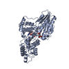 1dnpS S: Starting model for refinement |
|---|---|
| Similar structure data |


-Links
 PDBj
PDBj
- Assembly
| Deposited unit | 
| ||||||||
|---|---|---|---|---|---|---|---|---|---|
| 1 | 
| ||||||||
| 2 | 
| ||||||||
| Unit cell |
|
-Components
| #1: Protein | Mass: 57111.770 Da / Num. of mol.: 2 Source method: isolated from a genetically manipulated source Source: (gene. exp.) #2: Chemical |   Mass: 96.063 Da / Num. of mol.: 3 / Source method: obtained synthetically / Formula: SO4 Mass: 96.063 Da / Num. of mol.: 3 / Source method: obtained synthetically / Formula: SO4#3: Chemical |   Mass: 785.550 Da / Num. of mol.: 2 / Source method: obtained synthetically / Formula: C27H33N9O15P2 / Comment: FAD*YM Mass: 785.550 Da / Num. of mol.: 2 / Source method: obtained synthetically / Formula: C27H33N9O15P2 / Comment: FAD*YM#4: Water | ChemComp-HOH / |  Mass: 18.015 Da / Num. of mol.: 348 / Source method: isolated from a natural source / Formula: H2O Mass: 18.015 Da / Num. of mol.: 348 / Source method: isolated from a natural source / Formula: H2O |
|---|
-Experimental details
-Experiment
| Experiment | Method: X-RAY DIFFRACTION / Number of used crystals: 1 |
|---|
- Sample preparation
| Crystal | Density Matthews: 2.59 Å3/Da / Density % sol: 52.56 % | ||||||||||||||||||||||||||||||||||||||||||||||||||||||||
|---|---|---|---|---|---|---|---|---|---|---|---|---|---|---|---|---|---|---|---|---|---|---|---|---|---|---|---|---|---|---|---|---|---|---|---|---|---|---|---|---|---|---|---|---|---|---|---|---|---|---|---|---|---|---|---|---|---|
| Crystal grow | Temperature: 295 K / Method: vapor diffusion, hanging drop / pH: 6 Details: ammonium sulfate, pH 6.0, VAPOR DIFFUSION, HANGING DROP, temperature 295.0K | ||||||||||||||||||||||||||||||||||||||||||||||||||||||||
| Crystal grow | *PLUS Temperature: 20-24 ℃ / pH: 8 | ||||||||||||||||||||||||||||||||||||||||||||||||||||||||
| Components of the solutions | *PLUS
|
-Data collection
| Diffraction | Mean temperature: 100 K |
|---|---|
| Diffraction source | Source: SYNCHROTRON / Site: SSRL  / Beamline: BL7-1 / Wavelength: 1.08 Å / Beamline: BL7-1 / Wavelength: 1.08 Å |
| Detector | Type: MARRESEARCH / Detector: IMAGE PLATE / Date: Feb 3, 2000 |
| Radiation | Monochromator: Si(111) / Protocol: SINGLE WAVELENGTH / Monochromatic (M) / Laue (L): M / Scattering type: x-ray |
| Radiation wavelength | Wavelength: 1.08 Å / Relative weight: 1 |
| Reflection | Resolution: 1.9→30 Å / Num. all: 88628 / Num. obs: 88628 / % possible obs: 96.5 % / Observed criterion σ(F): 1 / Observed criterion σ(I): 1 / Redundancy: 2.6 % / Biso Wilson estimate: 22.7 Å2 / Rmerge(I) obs: 0.045 / Rsym value: 0.045 / Net I/σ(I): 16.5 |
| Reflection shell | Resolution: 1.9→1.97 Å / Redundancy: 1.6 % / Mean I/σ(I) obs: 2.9 / Num. unique all: 8731 / Rsym value: 0.311 / % possible all: 95.8 |
| Reflection | *PLUS Lowest resolution: 30 Å |
| Reflection shell | *PLUS % possible obs: 95.8 % / Rmerge(I) obs: 0.311 |
- Processing
| Software |
| ||||||||||||||||||||
|---|---|---|---|---|---|---|---|---|---|---|---|---|---|---|---|---|---|---|---|---|---|
| Refinement | Method to determine structure: MOLECULAR REPLACEMENT Starting model: PDB ENTRY 1DNP Resolution: 1.9→30 Å / Isotropic thermal model: Isotropic / Cross valid method: THROUGHOUT / σ(F): 1 / σ(I): 1 / Stereochemistry target values: Engh & Huber
| ||||||||||||||||||||
| Displacement parameters | Biso mean: 24 Å2 | ||||||||||||||||||||
| Refinement step | Cycle: LAST / Resolution: 1.9→30 Å
| ||||||||||||||||||||
| Refine LS restraints |
| ||||||||||||||||||||
| LS refinement shell | Resolution: 1.9→1.91 Å | ||||||||||||||||||||
| Refinement | *PLUS Lowest resolution: 30 Å | ||||||||||||||||||||
| Solvent computation | *PLUS | ||||||||||||||||||||
| Displacement parameters | *PLUS |
