 Movie
Movie Controller


+ Open data

- Basic information
| Entry | Database: PDB / ID: 1dnp | ||||||
|---|---|---|---|---|---|---|---|
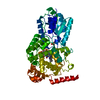
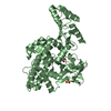
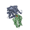
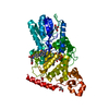
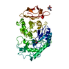
| Title | STRUCTURE OF DEOXYRIBODIPYRIMIDINE PHOTOLYASE | ||||||
 Components Components | DNA PHOTOLYASE | ||||||
 Keywords Keywords | LYASE (CARBON-CARBON) / DNA REPAIR / ELECTRON TRANSFER / EXCITATION ENERGY TRANSFER / LYASE / CARBON-CARBON | ||||||
| Function / homology |  Function and homology information Function and homology informationphotoreactive repair / deoxyribodipyrimidine photo-lyase / deoxyribodipyrimidine photo-lyase activity / phototransduction, visible light / response to light stimulus / FAD binding / damaged DNA binding / DNA binding Similarity search - Function | ||||||
| Biological species |  | ||||||
| Method |  X-RAY DIFFRACTION / Resolution: 2.3 Å X-RAY DIFFRACTION / Resolution: 2.3 Å | ||||||
 Authors Authors | Park, H.-W. / Sancar, A. / Deisenhofer, J. | ||||||
 Citation Citation | Journal: Science / Year: 1995 Title: Crystal structure of DNA photolyase from Escherichia coli. Authors: Park, H.W. / Kim, S.T. / Sancar, A. / Deisenhofer, J. #1: Journal: J.Mol.Biol. / Year: 1993Title: Crystallization and Preliminary Crystallographic Analysis of Escherichia Coli DNA Photolyase Authors: Park, H.W. / Sancar, A. / Deisenhofer, J. | ||||||
| History |
|
- Structure visualization
| Structure viewer | Molecule: MolmilJmol/JSmol |
|---|
- Downloads & links
-Download
| PDBx/mmCIF format | 1dnp.cif.gz | 205.4 KB | Display | PDBx/mmCIF format |
|---|---|---|---|---|
| PDB format | pdb1dnp.ent.gz | 164.2 KB | Display | PDB format |
| PDBx/mmJSON format | 1dnp.json.gz | Tree view | PDBx/mmJSON format | |
| Others |  Other downloads Other downloads |
-Validation report
| Arichive directory | https://data.pdbj.org/pub/pdb/validation_reports/dn/1dnpftp://data.pdbj.org/pub/pdb/validation_reports/dn/1dnp | HTTPS FTP |
|---|
-Related structure data
| Similar structure data |
|---|


-Links
 PDBj
PDBj
- Assembly
| Deposited unit | 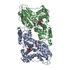
| ||||||||
|---|---|---|---|---|---|---|---|---|---|
| 1 | 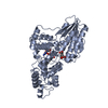
| ||||||||
| 2 | 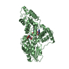
| ||||||||
| Unit cell |
| ||||||||
| Noncrystallographic symmetry (NCS) | NCS oper: (Code: given Matrix: (0.025044, -0.324306, -0.945621), Vector: |
-Components
| #1: Protein | Mass: 53601.562 Da / Num. of mol.: 2 / Source method: isolated from a natural source / Details: PHOTOREACTIVATING ENZYME / Source: (natural) References: UniProt: P00914, deoxyribodipyrimidine photo-lyase #2: Chemical |   Mass: 785.550 Da / Num. of mol.: 2 / Source method: obtained synthetically / Formula: C27H33N9O15P2 / Comment: FAD*YM Mass: 785.550 Da / Num. of mol.: 2 / Source method: obtained synthetically / Formula: C27H33N9O15P2 / Comment: FAD*YM#3: Chemical |   Mass: 457.440 Da / Num. of mol.: 2 / Source method: obtained synthetically / Formula: C20H23N7O6 Mass: 457.440 Da / Num. of mol.: 2 / Source method: obtained synthetically / Formula: C20H23N7O6#4: Water | ChemComp-HOH / |  Mass: 18.015 Da / Num. of mol.: 373 / Source method: isolated from a natural source / Formula: H2O Mass: 18.015 Da / Num. of mol.: 373 / Source method: isolated from a natural source / Formula: H2O |
|---|
-Experimental details
-Experiment
| Experiment | Method: X-RAY DIFFRACTION |
|---|
- Sample preparation
| Crystal | Density Matthews: 2.27 Å3/Da / Density % sol: 47 % | ||||||||||||||||||||||||||||||||||||||||||||||||
|---|---|---|---|---|---|---|---|---|---|---|---|---|---|---|---|---|---|---|---|---|---|---|---|---|---|---|---|---|---|---|---|---|---|---|---|---|---|---|---|---|---|---|---|---|---|---|---|---|---|
| Crystal | *PLUS | ||||||||||||||||||||||||||||||||||||||||||||||||
| Crystal grow | *PLUS pH: 7.6 / Method: vapor diffusionDetails: seeding. refer to Park, H.W., (1993) J.Mol.Biol., 231, 1122. | ||||||||||||||||||||||||||||||||||||||||||||||||
| Components of the solutions | *PLUS
|
-Data collection
| Diffraction source | Source: ROTATING ANODE / Wavelength: 1.5418 |
|---|---|
| Detector | Type: XUONG-HAMLIN MULTIWIRE / Detector: AREA DETECTOR / Date: Oct 1, 1991 |
| Radiation | Monochromatic (M) / Laue (L): M / Scattering type: x-ray |
| Radiation wavelength | Wavelength: 1.5418 Å / Relative weight: 1 |
| Reflection | Highest resolution: 2.3 Å / Num. obs: 33086 / % possible obs: 78.3 % / Observed criterion σ(I): 2 / Redundancy: 2.45 % / Rmerge(I) obs: 0.043 |
| Reflection | *PLUS Num. measured all: 76311 |
- Processing
| Software |
| ||||||||||||||||||||||||||||||||||||||||||||||||||||||||||||
|---|---|---|---|---|---|---|---|---|---|---|---|---|---|---|---|---|---|---|---|---|---|---|---|---|---|---|---|---|---|---|---|---|---|---|---|---|---|---|---|---|---|---|---|---|---|---|---|---|---|---|---|---|---|---|---|---|---|---|---|---|---|
| Refinement | Resolution: 2.3→10 Å / σ(F): 2
| ||||||||||||||||||||||||||||||||||||||||||||||||||||||||||||
| Displacement parameters | Biso mean: 17.62 Å2 | ||||||||||||||||||||||||||||||||||||||||||||||||||||||||||||
| Refine analyze | Luzzati sigma a obs: 0.27 Å | ||||||||||||||||||||||||||||||||||||||||||||||||||||||||||||
| Refinement step | Cycle: LAST / Resolution: 2.3→10 Å
| ||||||||||||||||||||||||||||||||||||||||||||||||||||||||||||
| Refine LS restraints |
| ||||||||||||||||||||||||||||||||||||||||||||||||||||||||||||
| Software | *PLUS Name: X-PLOR / Version: 3.1 / Classification: refinement | ||||||||||||||||||||||||||||||||||||||||||||||||||||||||||||
| Refine LS restraints | *PLUS
|
