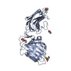
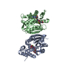
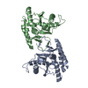
[English] 日本語
Yorodumi
- PDB-5ik5: Laminin A2LG45 C-form, G6/7 bound. -

+
Open data


ID or keywords:

Loading...

-
Basic information

Entry
Database: PDB / ID: 5ik5
TitleLaminin A2LG45 C-form, G6/7 bound.
ComponentsLaminin subunit alpha-2
KeywordsSTRUCTURAL PROTEIN / Extracellular Matrix / Ligand binding / LG domain
Function / homology
Function and homology information


regulation of basement membrane organization / Schwann cell differentiation / positive regulation of synaptic transmission, cholinergic / positive regulation of integrin-mediated signaling pathway / tissue development / protein complex involved in cell-matrix adhesion / extracellular matrix structural constituent / positive regulation of muscle cell differentiation / regulation of embryonic development / basement membrane ...regulation of basement membrane organization / Schwann cell differentiation / positive regulation of synaptic transmission, cholinergic / positive regulation of integrin-mediated signaling pathway / tissue development / protein complex involved in cell-matrix adhesion / extracellular matrix structural constituent / positive regulation of muscle cell differentiation / regulation of embryonic development / basement membrane / positive regulation of cell adhesion / synaptic cleft / regulation of cell migration / axon guidance / animal organ morphogenesis / sarcolemma / neuromuscular junction / collagen-containing extracellular matrix / dendritic spine / cell adhesion / signaling receptor binding / extracellular region
Similarity search - Function
Laminin alpha, domain I / Laminin domain II / Laminin Domain I / Laminin Domain II / Laminin IV / Laminin B (Domain IV) / Laminin IV type A domain profile. / Laminin B domain / Laminin, N-terminal / Laminin N-terminal (Domain VI) ...Laminin alpha, domain I / Laminin domain II / Laminin Domain I / Laminin Domain II / Laminin IV / Laminin B (Domain IV) / Laminin IV type A domain profile. / Laminin B domain / Laminin, N-terminal / Laminin N-terminal (Domain VI) / Laminin N-terminal domain profile. / Laminin N-terminal domain (domain VI) / Laminin-type EGF-like (LE) domain profile. / Laminin G domain / Laminin-type EGF-like (LE) domain signature. / Laminin-type epidermal growth factor-like domai / Laminin EGF domain / Laminin-type EGF domain / Laminin G domain / Laminin G domain profile. / Laminin G domain / Laminin G domain / Epidermal growth factor-like domain. / Jelly Rolls - #200 / Growth factor receptor cysteine-rich domain superfamily / EGF-like domain signature 2. / EGF-like domain signature 1. / EGF-like domain / Concanavalin A-like lectin/glucanase domain superfamily / Jelly Rolls / Sandwich / Mainly Beta
Similarity search - Domain/homology
Thomsen-Friedenreich antigen / 7-hydroxy-4-methyl-2H-chromen-2-one / Laminin subunit alpha-2
Similarity search - Component
Biological speciesMus musculus (house mouse)
MethodX-RAY DIFFRACTION / SYNCHROTRON / MOLECULAR REPLACEMENT / Resolution: 1.39 Å
AuthorsBriggs, D.C. / Hohenester, E. / Campbell, K.P.
Funding support United Kingdom, 1items
OrganizationGrant numberCountry
Wellcome Trust101748/Z/13/Z United Kingdom
CitationJournal: Nat.Chem.Biol. / Year: 2016
Title: Structural basis of laminin binding to the LARGE glycans on dystroglycan.
Authors: Briggs, D.C. / Yoshida-Moriguchi, T. / Zheng, T. / Venzke, D. / Anderson, M.E. / Strazzulli, A. / Moracci, M. / Yu, L. / Hohenester, E. / Campbell, K.P.
History
DepositionMar 3, 2016Deposition site: RCSB / Processing site: PDBE
Revision 1.0Aug 10, 2016Provider: repository / Type: Initial release
Revision 1.1Aug 24, 2016Group: Database references
Revision 1.2Sep 28, 2016Group: Database references
Revision 2.0Jul 29, 2020Group: Advisory / Atomic model ...Advisory / Atomic model / Data collection / Derived calculations / Structure summary
Category: atom_site / atom_site_anisotrop ...atom_site / atom_site_anisotrop / chem_comp / entity / entity_name_com / pdbx_branch_scheme / pdbx_chem_comp_identifier / pdbx_entity_branch / pdbx_entity_branch_descriptor / pdbx_entity_branch_link / pdbx_entity_branch_list / pdbx_entity_nonpoly / pdbx_molecule_features / pdbx_nonpoly_scheme / pdbx_struct_assembly_gen / pdbx_struct_conn_angle / pdbx_struct_special_symmetry / pdbx_validate_close_contact / struct_asym / struct_conn / struct_site / struct_site_gen
Item: _atom_site.B_iso_or_equiv / _atom_site.Cartn_x ..._atom_site.B_iso_or_equiv / _atom_site.Cartn_x / _atom_site.Cartn_y / _atom_site.Cartn_z / _atom_site.auth_asym_id / _atom_site.auth_atom_id / _atom_site.auth_comp_id / _atom_site.auth_seq_id / _atom_site.label_alt_id / _atom_site.label_asym_id / _atom_site.label_atom_id / _atom_site.label_comp_id / _atom_site.label_entity_id / _atom_site.occupancy / _atom_site.type_symbol / _atom_site_anisotrop.U[1][1] / _atom_site_anisotrop.U[1][2] / _atom_site_anisotrop.U[1][3] / _atom_site_anisotrop.U[2][2] / _atom_site_anisotrop.U[2][3] / _atom_site_anisotrop.U[3][3] / _atom_site_anisotrop.id / _atom_site_anisotrop.pdbx_auth_asym_id / _atom_site_anisotrop.pdbx_auth_atom_id / _atom_site_anisotrop.pdbx_auth_comp_id / _atom_site_anisotrop.pdbx_auth_seq_id / _atom_site_anisotrop.pdbx_label_asym_id / _atom_site_anisotrop.pdbx_label_atom_id / _atom_site_anisotrop.pdbx_label_comp_id / _atom_site_anisotrop.type_symbol / _chem_comp.name / _chem_comp.type / _pdbx_struct_assembly_gen.asym_id_list / _pdbx_struct_conn_angle.ptnr1_auth_asym_id / _pdbx_struct_conn_angle.ptnr1_auth_comp_id / _pdbx_struct_conn_angle.ptnr1_auth_seq_id / _pdbx_struct_conn_angle.ptnr1_label_alt_id / _pdbx_struct_conn_angle.ptnr1_label_asym_id / _pdbx_struct_conn_angle.ptnr1_label_atom_id / _pdbx_struct_conn_angle.ptnr1_label_comp_id / _pdbx_struct_conn_angle.ptnr1_label_seq_id / _pdbx_struct_conn_angle.ptnr2_label_asym_id / _pdbx_struct_conn_angle.ptnr2_symmetry / _pdbx_struct_conn_angle.ptnr3_auth_asym_id / _pdbx_struct_conn_angle.ptnr3_auth_comp_id / _pdbx_struct_conn_angle.ptnr3_auth_seq_id / _pdbx_struct_conn_angle.ptnr3_label_alt_id / _pdbx_struct_conn_angle.ptnr3_label_asym_id / _pdbx_struct_conn_angle.ptnr3_label_atom_id / _pdbx_struct_conn_angle.ptnr3_label_comp_id / _pdbx_struct_conn_angle.ptnr3_label_seq_id / _pdbx_struct_conn_angle.value / _pdbx_struct_special_symmetry.label_asym_id / _pdbx_validate_close_contact.auth_asym_id_1 / _pdbx_validate_close_contact.auth_asym_id_2 / _pdbx_validate_close_contact.auth_seq_id_1 / _pdbx_validate_close_contact.auth_seq_id_2 / _struct_conn.conn_type_id / _struct_conn.id / _struct_conn.pdbx_dist_value / _struct_conn.pdbx_leaving_atom_flag / _struct_conn.pdbx_ptnr1_label_alt_id / _struct_conn.pdbx_ptnr2_label_alt_id / _struct_conn.pdbx_role / _struct_conn.ptnr1_auth_asym_id / _struct_conn.ptnr1_auth_comp_id / _struct_conn.ptnr1_auth_seq_id / _struct_conn.ptnr1_label_asym_id / _struct_conn.ptnr1_label_atom_id / _struct_conn.ptnr1_label_comp_id / _struct_conn.ptnr1_label_seq_id / _struct_conn.ptnr2_auth_asym_id / _struct_conn.ptnr2_auth_comp_id / _struct_conn.ptnr2_auth_seq_id / _struct_conn.ptnr2_label_asym_id / _struct_conn.ptnr2_label_atom_id / _struct_conn.ptnr2_label_comp_id / _struct_conn.ptnr2_symmetry
Description: Carbohydrate remediation / Provider: repository / Type: Remediation
Revision 2.1Jan 10, 2024Group: Data collection / Database references ...Data collection / Database references / Derived calculations / Refinement description / Structure summary
Category: chem_comp / chem_comp_atom ...chem_comp / chem_comp_atom / chem_comp_bond / database_2 / pdbx_initial_refinement_model / struct_conn
Item: _chem_comp.pdbx_synonyms / _database_2.pdbx_DOI ..._chem_comp.pdbx_synonyms / _database_2.pdbx_DOI / _database_2.pdbx_database_accession / _struct_conn.pdbx_leaving_atom_flag

-
Structure visualization

Structure viewerMolecule:
MolmilJmol/JSmol

Downloads & links

-
Assembly

Deposited unit
A: Laminin subunit alpha-2
hetero molecules


Theoretical massNumber of molelcules
Total (without water)44,88013
Polymers42,6281
Non-polymers2,25112
Water8,287460
1


  • Idetical with deposited unit
  • defined by author&software
TypeNameSymmetry operationNumber
identity operation1_555x,y,z1
MethodPISA
Unit cell
Length a, b, c (Å)70.390, 110.740, 123.900
Angle α, β, γ (deg.)90.00, 90.00, 90.00
Int Tables number20
Space group name H-MC2221
Components on special symmetry positions
IDModelComponents
11A-4013-

4MU

21A-4533-

HOH

-
Components

-
Protein , 1 types, 1 molecules A

#1: Protein Laminin subunit alpha-2 / / Laminin M chain / Laminin-12 subunit alpha / Laminin-2 subunit alpha / Laminin-4 subunit alpha / ...Laminin M chain / Laminin-12 subunit alpha / Laminin-2 subunit alpha / Laminin-4 subunit alpha / Merosin heavy chain


Mass: 42628.438 Da / Num. of mol.: 1 / Fragment: UNP residues 2730-3118
Source method: isolated from a genetically manipulated source
Source: (gene. exp.) Mus musculus (house mouse) / Gene: Lama2 / Plasmid: pCEP-Pu / Cell line (production host): HEK293 / Production host: Homo sapiens (human) / References: UniProt: Q60675

-
Sugars , 3 types, 3 molecules

#2: Polysaccharide beta-D-galactopyranose-(1-3)-2-acetamido-2-deoxy-alpha-D-galactopyranose / Thomsen-Friedenreich antigen / Thomsen–Friedenreich antigen


Type: oligosaccharide, Oligosaccharide / Class: Antigen / Mass: 383.349 Da / Num. of mol.: 1
Source method: isolated from a genetically manipulated source
Details: oligosaccharide / References: Thomsen-Friedenreich antigen
DescriptorTypeProgram
DGalpb1-3DGalpNAca1-Glycam Condensed SequenceGMML 1.0
WURCS=2.0/2,2,1/[a2112h-1a_1-5_2*NCC/3=O][a2112h-1b_1-5]/1-2/a3-b1WURCSPDB2Glycan 1.1.0
[]{[(3+1)][a-D-GalpNAc]{[(3+1)][b-D-Galp]{}}}LINUCSPDB-CARE
#3: Polysaccharide 2-acetamido-2-deoxy-beta-D-glucopyranose-(1-4)-2-acetamido-2-deoxy-beta-D-glucopyranose


Type: oligosaccharide / Mass: 424.401 Da / Num. of mol.: 1
Source method: isolated from a genetically manipulated source
DescriptorTypeProgram
DGlcpNAcb1-4DGlcpNAcb1-Glycam Condensed SequenceGMML 1.0
WURCS=2.0/1,2,1/[a2122h-1b_1-5_2*NCC/3=O]/1-1/a4-b1WURCSPDB2Glycan 1.1.0
[]{[(4+1)][b-D-GlcpNAc]{[(4+1)][b-D-GlcpNAc]{}}}LINUCSPDB-CARE
#4: Polysaccharide alpha-D-xylopyranose-(1-3)-beta-D-glucopyranuronic acid-(1-3)-alpha-D-xylopyranose-(1-3)-beta-D- ...alpha-D-xylopyranose-(1-3)-beta-D-glucopyranuronic acid-(1-3)-alpha-D-xylopyranose-(1-3)-beta-D-glucopyranuronic acid


Type: oligosaccharide / Mass: 634.492 Da / Num. of mol.: 1
Source method: isolated from a genetically manipulated source
DescriptorTypeProgram
DXylpa1-3DGlcpAb1-3DXylpa1-3DGlcpAb1-ROHGlycam Condensed SequenceGMML 1.0
WURCS=2.0/2,4,3/[a2122A-1b_1-5][a212h-1a_1-5]/1-2-1-2/a3-b1_b3-c1_c3-d1WURCSPDB2Glycan 1.1.0
[][D-1-deoxy-GlcpA]{[(3+1)][a-D-Xylp]{[(3+1)][b-D-GlcpA]{[(3+1)][a-D-Xylp]{}}}}LINUCSPDB-CARE

-
Non-polymers , 4 types, 469 molecules

#5: Chemical ChemComp-CA / CALCIUM ION


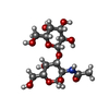
Mass: 40.078 Da / Num. of mol.: 2 / Source method: obtained synthetically / Formula: Ca
#6: Chemical
ChemComp-GOL / GLYCEROL / GLYCERIN / PROPANE-1,2,3-TRIOL / Glycerol


Mass: 92.094 Da / Num. of mol.: 6 / Source method: obtained synthetically / Formula: C3H8O3
#7: Chemical ChemComp-4MU / 7-hydroxy-4-methyl-2H-chromen-2-one / 4-methylumbelliferone / Hymecromone


Mass: 176.169 Da / Num. of mol.: 1 / Source method: obtained synthetically / Formula: C10H8O3
#8: Water ChemComp-HOH / water / Water


Mass: 18.015 Da / Num. of mol.: 460 / Source method: isolated from a natural source / Formula: H2O

-
Experimental details

-
Experiment

ExperimentMethod: X-RAY DIFFRACTION / Number of used crystals: 1

-
Sample preparation

CrystalDensity Matthews: 2.83 Å3/Da / Density % sol: 56.57 %
Crystal growTemperature: 292 K / Method: vapor diffusion, hanging drop / pH: 7.5
Details: 100mM HEPES/MOPS 120mM Glucuronic Acid 30% PEG8k/Glycerol mix

-
Data collection

DiffractionMean temperature: 100 K
Diffraction sourceSource: SYNCHROTRON / Site: Diamond / Beamline: I02 / Wavelength: 0.9795 Å
DetectorType: DECTRIS PILATUS 6M-F / Detector: PIXEL / Date: May 14, 2015
RadiationProtocol: SINGLE WAVELENGTH / Monochromatic (M) / Laue (L): M / Scattering type: x-ray
Radiation wavelengthWavelength: 0.9795 Å / Relative weight: 1
ReflectionResolution: 1.39→61.95 Å / Num. obs: 81183 / % possible obs: 83.7 % / Redundancy: 3.9 % / Biso Wilson estimate: 18.04 Å2 / CC1/2: 0.999 / Rmerge(I) obs: 0.035 / Net I/σ(I): 17.3
Reflection shellResolution: 1.39→1.44 Å / Redundancy: 1.6 % / Rmerge(I) obs: 0.422 / Mean I/σ(I) obs: 1.3 / % possible all: 35.8

-
Processing

Software
NameVersionClassification
PHENIX(1.10.1_2155: ???)refinement
XDSdata reduction
Aimlessdata scaling
PHASERphasing
RefinementMethod to determine structure: MOLECULAR REPLACEMENT
Starting model: 1DYK
Resolution: 1.39→61.95 Å / SU ML: 0.13 / Cross valid method: FREE R-VALUE / σ(F): 1.34 / Phase error: 22.15 / Stereochemistry target values: ML
RfactorNum. reflection% reflectionSelection details
Rfree0.1768 4052 4.99 %Random Selection
Rwork0.1489 ---
obs0.1504 81182 83.5 %-
Solvent computationShrinkage radii: 0.9 Å / VDW probe radii: 1.11 Å / Solvent model: FLAT BULK SOLVENT MODEL
Refinement stepCycle: LAST / Resolution: 1.39→61.95 Å
ProteinNucleic acidLigandSolventTotal
Num. atoms2888 0 146 460 3494
Refine LS restraints
Refine-IDTypeDev idealNumber
X-RAY DIFFRACTIONf_bond_d0.0123182
X-RAY DIFFRACTIONf_angle_d0.8954310
X-RAY DIFFRACTIONf_dihedral_angle_d15.2241204
X-RAY DIFFRACTIONf_chiral_restr0.079503
X-RAY DIFFRACTIONf_plane_restr0.005543
LS refinement shell
Resolution (Å)Rfactor RfreeNum. reflection RfreeRfactor RworkNum. reflection RworkRefine-ID% reflection obs (%)
1.39-1.40640.305500.2633995X-RAY DIFFRACTION32
1.4064-1.42350.2825510.25491221X-RAY DIFFRACTION38
1.4235-1.44150.2936700.23271325X-RAY DIFFRACTION42
1.4415-1.46050.3073630.22571548X-RAY DIFFRACTION48
1.4605-1.48050.2711780.20961659X-RAY DIFFRACTION52
1.4805-1.50170.26481030.19931817X-RAY DIFFRACTION58
1.5017-1.52410.24581010.19321960X-RAY DIFFRACTION62
1.5241-1.54790.22411200.18842112X-RAY DIFFRACTION68
1.5479-1.57330.22961290.16982326X-RAY DIFFRACTION74
1.5733-1.60040.22261270.15892519X-RAY DIFFRACTION78
1.6004-1.62950.24781350.16012684X-RAY DIFFRACTION86
1.6295-1.66090.19821410.15582882X-RAY DIFFRACTION91
1.6609-1.69480.19271440.15723055X-RAY DIFFRACTION96
1.6948-1.73160.19941710.15163155X-RAY DIFFRACTION100
1.7316-1.77190.20921940.153135X-RAY DIFFRACTION100
1.7719-1.81620.19171320.14833211X-RAY DIFFRACTION100
1.8162-1.86530.18431630.14283167X-RAY DIFFRACTION100
1.8653-1.92020.20211550.14183165X-RAY DIFFRACTION100
1.9202-1.98220.16061850.13833150X-RAY DIFFRACTION99
1.9822-2.05310.13931620.13523177X-RAY DIFFRACTION100
2.0531-2.13530.18411500.13623188X-RAY DIFFRACTION100
2.1353-2.23240.15911770.13483163X-RAY DIFFRACTION100
2.2324-2.35010.16261600.14573205X-RAY DIFFRACTION100
2.3501-2.49740.17041560.14463176X-RAY DIFFRACTION99
2.4974-2.69020.18771480.15573221X-RAY DIFFRACTION100
2.6902-2.96090.18911780.14923211X-RAY DIFFRACTION100
2.9609-3.38940.18821930.14623166X-RAY DIFFRACTION99
3.3894-4.27010.1521980.13333223X-RAY DIFFRACTION99
4.2701-62.01160.16192180.16033314X-RAY DIFFRACTION99

+
About Yorodumi

-
News

-
Feb 9, 2022. New format data for meta-information of EMDB entries

New format data for meta-information of EMDB entries

  • Version 3 of the EMDB header file is now the official format.
  • The previous official version 1.9 will be removed from the archive.

Related info.:EMDB header

External links:wwPDB to switch to version 3 of the EMDB data model

-
Aug 12, 2020. Covid-19 info

Covid-19 info

URL: https://pdbj.org/emnavi/covid19.php

New page: Covid-19 featured information page in EM Navigator.

Related info.:Covid-19 info / Mar 5, 2020. Novel coronavirus structure data

+
Mar 5, 2020. Novel coronavirus structure data

Novel coronavirus structure data

Related info.:Yorodumi Speices / Aug 12, 2020. Covid-19 info

External links:COVID-19 featured content - PDBj / Molecule of the Month (242):Coronavirus Proteases

+
Jan 31, 2019. EMDB accession codes are about to change! (news from PDBe EMDB page)

EMDB accession codes are about to change! (news from PDBe EMDB page)

  • The allocation of 4 digits for EMDB accession codes will soon come to an end. Whilst these codes will remain in use, new EMDB accession codes will include an additional digit and will expand incrementally as the available range of codes is exhausted. The current 4-digit format prefixed with “EMD-” (i.e. EMD-XXXX) will advance to a 5-digit format (i.e. EMD-XXXXX), and so on. It is currently estimated that the 4-digit codes will be depleted around Spring 2019, at which point the 5-digit format will come into force.
  • The EM Navigator/Yorodumi systems omit the EMD- prefix.

Related info.:Q: What is EMD? / ID/Accession-code notation in Yorodumi/EM Navigator

External links:EMDB Accession Codes are Changing Soon! / Contact to PDBj

+
Jul 12, 2017. Major update of PDB

Major update of PDB

  • wwPDB released updated PDB data conforming to the new PDBx/mmCIF dictionary.
  • This is a major update changing the version number from 4 to 5, and with Remediation, in which all the entries are updated.
  • In this update, many items about electron microscopy experimental information are reorganized (e.g. em_software).
  • Now, EM Navigator and Yorodumi are based on the updated data.

External links:wwPDB Remediation / Enriched Model Files Conforming to OneDep Data Standards Now Available in the PDB FTP Archive

-
Yorodumi

Thousand views of thousand structures

  • Yorodumi is a browser for structure data from EMDB, PDB, SASBDB, etc.
  • This page is also the successor to EM Navigator detail page, and also detail information page/front-end page for Omokage search.
  • The word "yorodu" (or yorozu) is an old Japanese word meaning "ten thousand". "mi" (miru) is to see.

Related info.:EMDB / PDB / SASBDB / Comparison of 3 databanks / Yorodumi Search / Aug 31, 2016. New EM Navigator & Yorodumi / Yorodumi Papers / Jmol/JSmol / Function and homology information / Changes in new EM Navigator and Yorodumi

Read more