 Movie
Movie Controller

[English] 日本語
 Yorodumi
- PDB-5ii2: Crystal Structure of the fifth bromodomain of human polybromo (PB... -
+ Open data

- Basic information
| Entry | Database: PDB / ID: 5ii2 | ||||||
|---|---|---|---|---|---|---|---|
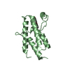
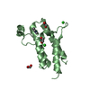
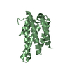
| Title | Crystal Structure of the fifth bromodomain of human polybromo (PB1) in complex with 2-(3,4-dihydroxyphenyl)-5,7-dihydroxy-4H-chromen-4-one | ||||||
 Components Components | Protein polybromo-1 | ||||||
 Keywords Keywords | TRANSCRIPTION / bromodomain / complex / small molecule / structural genomics consortium / SGC | ||||||
| Function / homology |  Function and homology information Function and homology informationFormation of the polybromo-BAF (pBAF) complex / regulation of G0 to G1 transition / RSC-type complex / regulation of nucleotide-excision repair / regulation of mitotic metaphase/anaphase transition / SWI/SNF complex / positive regulation of T cell differentiation / nuclear chromosome / positive regulation of double-strand break repair / RUNX1 interacts with co-factors whose precise effect on RUNX1 targets is not known ...Formation of the polybromo-BAF (pBAF) complex / regulation of G0 to G1 transition / RSC-type complex / regulation of nucleotide-excision repair / regulation of mitotic metaphase/anaphase transition / SWI/SNF complex / positive regulation of T cell differentiation / nuclear chromosome / positive regulation of double-strand break repair / RUNX1 interacts with co-factors whose precise effect on RUNX1 targets is not known / positive regulation of myoblast differentiation / regulation of G1/S transition of mitotic cell cycle / positive regulation of cell differentiation / transcription elongation by RNA polymerase II / kinetochore / nuclear matrix / RMTs methylate histone arginines / mitotic cell cycle / chromatin remodeling / negative regulation of cell population proliferation / chromatin binding / regulation of transcription by RNA polymerase II / chromatin / DNA binding / nucleoplasm / nucleus Similarity search - Function | ||||||
| Biological species |  Homo sapiens (human) Homo sapiens (human) | ||||||
| Method |  X-RAY DIFFRACTION / MOLECULAR REPLACEMENT / molecular replacement / Resolution: 2.1 Å X-RAY DIFFRACTION / MOLECULAR REPLACEMENT / molecular replacement / Resolution: 2.1 Å | ||||||
 Authors Authors | Filippakopoulos, P. / Picaud, S. / Felletar, I. / von Delft, F. / Edwards, A.M. / Arrowsmith, C.H. / Bountra, C. / Knapp, S. | ||||||
| Funding support |  United Kingdom, 1items United Kingdom, 1items
| ||||||
 Citation Citation | Journal: J.Med.Chem. / Year: 2016 Title: Discovery and Optimization of a Selective Ligand for the Switch/Sucrose Nonfermenting-Related Bromodomains of Polybromo Protein-1 by the Use of Virtual Screening and Hydration Analysis. Authors: Myrianthopoulos, V. / Gaboriaud-Kolar, N. / Tallant, C. / Hall, M.L. / Grigoriou, S. / Brownlee, P.M. / Fedorov, O. / Rogers, C. / Heidenreich, D. / Wanior, M. / Drosos, N. / Mexia, N. / ...Authors: Myrianthopoulos, V. / Gaboriaud-Kolar, N. / Tallant, C. / Hall, M.L. / Grigoriou, S. / Brownlee, P.M. / Fedorov, O. / Rogers, C. / Heidenreich, D. / Wanior, M. / Drosos, N. / Mexia, N. / Savitsky, P. / Bagratuni, T. / Kastritis, E. / Terpos, E. / Filippakopoulos, P. / Muller, S. / Skaltsounis, A.L. / Downs, J.A. / Knapp, S. / Mikros, E. | ||||||
| History |
|
- Structure visualization
| Structure viewer | Molecule: MolmilJmol/JSmol |
|---|
- Downloads & links
-Download
| PDBx/mmCIF format | 5ii2.cif.gz | 112.2 KB | Display | PDBx/mmCIF format |
|---|---|---|---|---|
| PDB format | pdb5ii2.ent.gz | 85.7 KB | Display | PDB format |
| PDBx/mmJSON format | 5ii2.json.gz | Tree view | PDBx/mmJSON format | |
| Others |  Other downloads Other downloads |
-Validation report
| Arichive directory | https://data.pdbj.org/pub/pdb/validation_reports/ii/5ii2ftp://data.pdbj.org/pub/pdb/validation_reports/ii/5ii2 | HTTPS FTP |
|---|
-Related structure data
| Related structure data |  5hrvC  5hrwC  5hrxC  5ii1C  5iidC 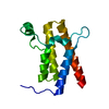 2grcS 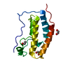 2ossS 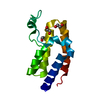 2ouoS  3d7cS  3daiS  3dwyS  3hmhS  3mb4S S: Starting model for refinement C: citing same article ( |
|---|---|
| Similar structure data |


-Links
 PDBj
PDBj


- Assembly
| Deposited unit | 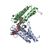
| ||||||||
|---|---|---|---|---|---|---|---|---|---|
| 1 | 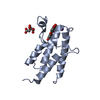
| ||||||||
| 2 | 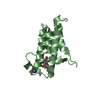
| ||||||||
| Unit cell |
|
-Components
| #1: Protein | Mass: 14648.000 Da / Num. of mol.: 2 Source method: isolated from a genetically manipulated source Source: (gene. exp.) Homo sapiens (human) / Gene: PBRM1, BAF180, PB1 / Plasmid: pNIC28-Bsa4 / Production host:  #2: Chemical |   Mass: 39.098 Da / Num. of mol.: 2 / Source method: obtained synthetically / Formula: K Mass: 39.098 Da / Num. of mol.: 2 / Source method: obtained synthetically / Formula: K#3: Chemical | ChemComp-CIT / |   Mass: 192.124 Da / Num. of mol.: 1 / Source method: obtained synthetically / Formula: C6H8O7 Mass: 192.124 Da / Num. of mol.: 1 / Source method: obtained synthetically / Formula: C6H8O7#4: Chemical |   Mass: 286.236 Da / Num. of mol.: 2 / Source method: obtained synthetically / Formula: C15H10O6 Mass: 286.236 Da / Num. of mol.: 2 / Source method: obtained synthetically / Formula: C15H10O6#5: Water | ChemComp-HOH / |  Mass: 18.015 Da / Num. of mol.: 182 / Source method: isolated from a natural source / Formula: H2O Mass: 18.015 Da / Num. of mol.: 182 / Source method: isolated from a natural source / Formula: H2O |
|---|
-Experimental details
-Experiment
| Experiment | Method: X-RAY DIFFRACTION / Number of used crystals: 1 |
|---|
- Sample preparation
| Crystal | Density Matthews: 2.87 Å3/Da / Density % sol: 57.08 % |
|---|---|
| Crystal grow | Temperature: 277 K / Method: vapor diffusion, sitting drop / pH: 7.5 Details: 0.20M K3(cit) 0.1M BTProp pH 7.5 20.0% PEG 3350 10.0% EtGly |
-Data collection
| Diffraction | Mean temperature: 100 K | ||||||||||||||||||||||||||||||||||||||||||||||||||||||||||||||||||
|---|---|---|---|---|---|---|---|---|---|---|---|---|---|---|---|---|---|---|---|---|---|---|---|---|---|---|---|---|---|---|---|---|---|---|---|---|---|---|---|---|---|---|---|---|---|---|---|---|---|---|---|---|---|---|---|---|---|---|---|---|---|---|---|---|---|---|---|
| Diffraction source | Source: ROTATING ANODE / Type: RIGAKU FR-E SUPERBRIGHT / Wavelength: 1.52 Å | ||||||||||||||||||||||||||||||||||||||||||||||||||||||||||||||||||
| Detector | Type: RIGAKU RAXIS IV / Detector: IMAGE PLATE / Date: Apr 5, 2011 | ||||||||||||||||||||||||||||||||||||||||||||||||||||||||||||||||||
| Radiation | Protocol: SINGLE WAVELENGTH / Monochromatic (M) / Laue (L): M / Scattering type: x-ray | ||||||||||||||||||||||||||||||||||||||||||||||||||||||||||||||||||
| Radiation wavelength | Wavelength: 1.52 Å / Relative weight: 1 | ||||||||||||||||||||||||||||||||||||||||||||||||||||||||||||||||||
| Reflection | Resolution: 2.1→24.186 Å / Num. obs: 20237 / % possible obs: 99 % / Redundancy: 6.3 % / Rmerge(I) obs: 0.127 / Rsym value: 0.127 / Net I/av σ(I): 3.929 / Net I/σ(I): 9.1 | ||||||||||||||||||||||||||||||||||||||||||||||||||||||||||||||||||
| Reflection shell |
|
-Phasing
| Phasing | Method: molecular replacement | |||||||||
|---|---|---|---|---|---|---|---|---|---|---|
| Phasing MR | Model details: Phaser MODE: MR_AUTO
|
- Processing
| Software |
| |||||||||||||||||||||||||||||||||||||||||||||||||||||||||||||||||||||||||||
|---|---|---|---|---|---|---|---|---|---|---|---|---|---|---|---|---|---|---|---|---|---|---|---|---|---|---|---|---|---|---|---|---|---|---|---|---|---|---|---|---|---|---|---|---|---|---|---|---|---|---|---|---|---|---|---|---|---|---|---|---|---|---|---|---|---|---|---|---|---|---|---|---|---|---|---|---|
| Refinement | Method to determine structure: MOLECULAR REPLACEMENT Starting model: Ensemble of 3MB4,3DAI,3HMH,2GRC,2OSS,2OUO,3D7C,3DWY Resolution: 2.1→24.186 Å / Cor.coef. Fo:Fc: 0.951 / Cor.coef. Fo:Fc free: 0.932 / SU B: 7.205 / SU ML: 0.105 / SU R Cruickshank DPI: 0.1658 / Cross valid method: THROUGHOUT / σ(F): 0 / ESU R: 0.166 / ESU R Free: 0.162 Details: HYDROGENS HAVE BEEN ADDED IN THE RIDING POSITIONS U VALUES : WITH TLS ADDED
| |||||||||||||||||||||||||||||||||||||||||||||||||||||||||||||||||||||||||||
| Solvent computation | Ion probe radii: 0.8 Å / Shrinkage radii: 0.8 Å / VDW probe radii: 1.2 Å | |||||||||||||||||||||||||||||||||||||||||||||||||||||||||||||||||||||||||||
| Displacement parameters | Biso max: 82.33 Å2 / Biso mean: 28.073 Å2 / Biso min: 14.17 Å2
| |||||||||||||||||||||||||||||||||||||||||||||||||||||||||||||||||||||||||||
| Refinement step | Cycle: final / Resolution: 2.1→24.186 Å
| |||||||||||||||||||||||||||||||||||||||||||||||||||||||||||||||||||||||||||
| Refine LS restraints |
| |||||||||||||||||||||||||||||||||||||||||||||||||||||||||||||||||||||||||||
| LS refinement shell | Resolution: 2.1→2.154 Å / Total num. of bins used: 20
| |||||||||||||||||||||||||||||||||||||||||||||||||||||||||||||||||||||||||||
| Refinement TLS params. | Method: refined / Refine-ID: X-RAY DIFFRACTION
| |||||||||||||||||||||||||||||||||||||||||||||||||||||||||||||||||||||||||||
| Refinement TLS group |
|
