[English] 日本語
 Yorodumi
- PDB-5i8f: Crystal structure of St. John's wort Hyp-1 protein in complex wit... -
+ Open data
- Basic information
| Entry | Database: PDB / ID: 5i8f | |||||||||
|---|---|---|---|---|---|---|---|---|---|---|
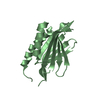
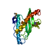
| Title | Crystal structure of St. John's wort Hyp-1 protein in complex with melatonin | |||||||||
 Components Components | Phenolic oxidative coupling protein | |||||||||
 Keywords Keywords | PLANT PROTEIN / PLANT HORMONE BINDING / PHYTOHORMONE BINDING / MELATONIN / CYTOKININ / PLANT DEFENSE / PATHOGENESIS-RELATED PROTEIN / PR-10 PROTEIN / HYPERICIN / DEPRESSION / PR-10 FOLD / HYDROPHOBIC CAVITY / ANS DISPLACEMENT ASSAY (ADA) | |||||||||
| Function / homology |  Function and homology information Function and homology informationabscisic acid binding / abscisic acid-activated signaling pathway / protein phosphatase inhibitor activity / defense response / signaling receptor activity / nucleus / cytoplasm Similarity search - Function | |||||||||
| Biological species |  Hypericum perforatum (plant) Hypericum perforatum (plant) | |||||||||
| Method |  X-RAY DIFFRACTION / X-RAY DIFFRACTION /  SYNCHROTRON / SYNCHROTRON /  MOLECULAR REPLACEMENT / Resolution: 1.3 Å MOLECULAR REPLACEMENT / Resolution: 1.3 Å | |||||||||
 Authors Authors | Sliwiak, J. / Dauter, Z. / Jaskolski, M. | |||||||||
| Funding support |  United States, United States,  Poland, 2items Poland, 2items
| |||||||||
 Citation Citation |  Journal: Front Plant Sci / Year: 2016 Journal: Front Plant Sci / Year: 2016Title: Crystal Structure of Hyp-1, a Hypericum perforatum PR-10 Protein, in Complex with Melatonin. Authors: Sliwiak, J. / Dauter, Z. / Jaskolski, M. #1:  Journal: J.Struct.Biol. / Year: 2010 Journal: J.Struct.Biol. / Year: 2010Title: Crystal Structure Of Hyp-1, A St. John'S Wort Protein Implicated In The Biosynthesis Of Hypericin. Authors: Michalska, K. / Fernandes, H. / Sikorski, M. / Jaskolski, M. #2: Journal: Acta Crystallogr.,Sect.D / Year: 2015 Title: Ans Complex Of St John'S Wort Pr-10 Protein With 28 Copies In The Asymmetric Unit: A Fiendish Combination Of Pseudosymmetry With Tetartohedral Twinning. Authors: Sliwiak, J. / Dauter, Z. / Kowiel, M. / Mccoy, A.J. / Read, R.J. / Jaskolski, M. #3:  Journal: Acta Crystallogr.,Sect.D / Year: 2014 Journal: Acta Crystallogr.,Sect.D / Year: 2014Title: Likelihood-Based Molecular-Replacement Solution For A Highly Pathological Crystal With Tetartohedral Twinning And Sevenfold Translational Noncrystallographic Symmetry. Authors: Sliwiak, J. / Jaskolski, M. / Dauter, Z. / Mccoy, A.J. / Read, R.J. #4:  Journal: J.Mol.Biol. / Year: 2002 Journal: J.Mol.Biol. / Year: 2002Title: Crystal Structures Of Two Homologous Pathogenesis-Related Proteins From Yellow Lupine. Authors: Biesiadka, J. / Bujacz, G. / Sikorski, M.M. / Jaskolski, M. #5:  Journal: J.Mol.Biol. / Year: 2008 Journal: J.Mol.Biol. / Year: 2008Title: Lupinus Luteus Pathogenesis-Related Protein As A Reservoir For Cytokinin. Authors: Fernandes, H. / Pasternak, O. / Bujacz, G. / Bujacz, A. / Sikorski, M.M. / Jaskolski, M. #6:  Journal: Febs J. / Year: 2009 Journal: Febs J. / Year: 2009Title: Cytokinin-Induced Structural Adaptability Of A Lupinus Luteus Pr-10 Protein. Authors: Fernandes, H. / Bujacz, A. / Bujacz, G. / Jelen, F. / Jasinski, M. / Kachlicki, P. / Otlewski, J. / Sikorski, M.M. / Jaskolski, M. #7:  Journal: Plant Cell / Year: 2006 Journal: Plant Cell / Year: 2006Title: Crystal Structure Of Vigna Radiata Cytokinin-Specific Binding Protein In Complex With Zeatin. Authors: Pasternak, O. / Bujacz, G.D. / Fujimoto, Y. / Hashimoto, Y. / Jelen, F. / Otlewski, J. / Sikorski, M.M. / Jaskolski, M. #8:  Journal: Acta Crystallogr.,Sect.D / Year: 2013 Journal: Acta Crystallogr.,Sect.D / Year: 2013Title: The Landscape Of Cytokinin Binding By A Plant Nodulin. Authors: Ruszkowski, M. / Szpotkowski, K. / Sikorski, M. / Jaskolski, M. #9:  Journal: Acta Crystallogr.,Sect.D / Year: 2014 Journal: Acta Crystallogr.,Sect.D / Year: 2014Title: Specific Binding Of Gibberellic Acid By Cytokinin-Specific Binding Proteins: A New Aspect Of Plant Hormone-Binding Proteins With The Pr-10 Fold. Authors: Ruszkowski, M. / Sliwiak, J. / Ciesielska, A. / Barciszewski, J. / Sikorski, M. / Jaskolski, M. #10:  Journal: J.Struct.Biol. / Year: 2016 Journal: J.Struct.Biol. / Year: 2016Title: Crystallographic And Cd Probing Of Ligand-Induced Conformational Changes In A Plant Pr-10 Protein. Authors: Sliwiak, J. / Dolot, R. / Michalska, K. / Szpotkowski, K. / Bujacz, G. / Sikorski, M. / Jaskolski, M. | |||||||||
| History |
|
- Structure visualization
| Structure viewer | Molecule:  Molmil Molmil Jmol/JSmol Jmol/JSmol |
|---|
- Downloads & links
- Download
| PDBx/mmCIF format |  5i8f.cif.gz 5i8f.cif.gz | 99.2 KB | Display |  PDBx/mmCIF format PDBx/mmCIF format |
|---|---|---|---|---|
| PDB format |  pdb5i8f.ent.gz pdb5i8f.ent.gz | 74.9 KB | Display |  PDB format PDB format |
| PDBx/mmJSON format |  5i8f.json.gz 5i8f.json.gz | Tree view |  PDBx/mmJSON format PDBx/mmJSON format | |
| Others |  Other downloads Other downloads |
-Validation report
| Summary document |  5i8f_validation.pdf.gz 5i8f_validation.pdf.gz | 446.9 KB | Display |  wwPDB validaton report wwPDB validaton report |
|---|---|---|---|---|
| Full document |  5i8f_full_validation.pdf.gz 5i8f_full_validation.pdf.gz | 448.9 KB | Display | |
| Data in XML |  5i8f_validation.xml.gz 5i8f_validation.xml.gz | 11.6 KB | Display | |
| Data in CIF |  5i8f_validation.cif.gz 5i8f_validation.cif.gz | 16.9 KB | Display | |
| Arichive directory |  https://data.pdbj.org/pub/pdb/validation_reports/i8/5i8f https://data.pdbj.org/pub/pdb/validation_reports/i8/5i8f ftp://data.pdbj.org/pub/pdb/validation_reports/i8/5i8f ftp://data.pdbj.org/pub/pdb/validation_reports/i8/5i8f | HTTPS FTP |
-Related structure data
| Related structure data |  3ie5S S: Starting model for refinement |
|---|---|
| Similar structure data | |
| Experimental dataset #1 | Data reference:  10.18150/repod.4711822 / Data set type: diffraction image data 10.18150/repod.4711822 / Data set type: diffraction image dataDetails: X-Ray synchrotron diffraction data for the crystal of Hypericum perforatum Hyp-1 protein in complex with melatonin |
- Links
Links
- Assembly
| Deposited unit | 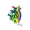
| ||||||||
|---|---|---|---|---|---|---|---|---|---|
| 1 |
| ||||||||
| Unit cell |
|
- Components
-Protein , 1 types, 1 molecules A
| #1: Protein | Mass: 18495.109 Da / Num. of mol.: 1 Source method: isolated from a genetically manipulated source Source: (gene. exp.)  Hypericum perforatum (plant) / Gene: hyp1 / Plasmid: PET151/D / Production host: Hypericum perforatum (plant) / Gene: hyp1 / Plasmid: PET151/D / Production host:  |
|---|
-Non-polymers , 5 types, 214 molecules 
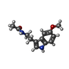


| #2: Chemical | ChemComp-UNL / Num. of mol.: 6 / Source method: obtained synthetically #3: Chemical | #4: Chemical | #5: Chemical | #6: Water | ChemComp-HOH / | |
|---|
-Experimental details
-Experiment
| Experiment | Method:  X-RAY DIFFRACTION / Number of used crystals: 1 X-RAY DIFFRACTION / Number of used crystals: 1 |
|---|
- Sample preparation
| Crystal | Density Matthews: 2.82 Å3/Da / Density % sol: 56.4 % / Description: prismatic |
|---|---|
| Crystal grow | Temperature: 292 K / Method: vapor diffusion, hanging drop / pH: 6.5 / Details: 1 M citrate (pH 6.5), 20% glycerol. |
-Data collection
| Diffraction | Mean temperature: 100 K |
|---|---|
| Diffraction source | Source:  SYNCHROTRON / Site: SYNCHROTRON / Site:  APS APS  / Beamline: 22-ID / Wavelength: 1 Å / Beamline: 22-ID / Wavelength: 1 Å |
| Detector | Type: MARMOSAIC 225 mm CCD / Detector: CCD / Date: Nov 21, 2011 |
| Radiation | Protocol: SINGLE WAVELENGTH / Monochromatic (M) / Laue (L): M / Scattering type: x-ray |
| Radiation wavelength | Wavelength: 1 Å / Relative weight: 1 |
| Reflection | Resolution: 1.3→30 Å / Num. obs: 51868 / % possible obs: 99.9 % / Redundancy: 2.6 % / Rmerge(I) obs: 0.057 / Net I/av σ(I): 28.9 / Net I/σ(I): 17 |
| Reflection shell | Resolution: 1.3→1.32 Å / Redundancy: 2 % / Rmerge(I) obs: 0.51 / Mean I/σ(I) obs: 2.5 / % possible all: 99.6 |
- Processing
| Software |
| ||||||||||||||||
|---|---|---|---|---|---|---|---|---|---|---|---|---|---|---|---|---|---|
| Refinement | Method to determine structure:  MOLECULAR REPLACEMENT MOLECULAR REPLACEMENTStarting model: 3ie5 Resolution: 1.3→30 Å / Cross valid method: FREE R-VALUE Details: Anisotropic refinement. H atoms were added at riding positions.
| ||||||||||||||||
| Refinement step | Cycle: LAST / Resolution: 1.3→30 Å
| ||||||||||||||||
| Refine LS restraints |
|
 Movie
Movie Controller





 PDBj
PDBj


