 Movie
Movie Controller

[English] 日本語
 Yorodumi
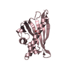
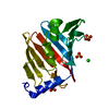
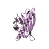
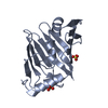
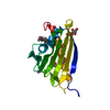
- PDB-1iwn: Crystal Structure of the Outer Membrane Lipoprotein Receptor LolB... -
+ Open data

- Basic information
| Entry | Database: PDB / ID: 1iwn | ||||||
|---|---|---|---|---|---|---|---|
| Title | Crystal Structure of the Outer Membrane Lipoprotein Receptor LolB Complexed with PEGMME2000 | ||||||
 Components Components | Outer Membrane Lipoprotein LolB | ||||||
 Keywords Keywords | PROTEIN TRANSPORT / UNCLOSED BETA BARREL / LIPOPROTEIN | ||||||
| Function / homology |  Function and homology information Function and homology informationlipoprotein localization to outer membrane / lipoprotein metabolic process / cell outer membrane / protein transport / outer membrane-bounded periplasmic space Similarity search - Function | ||||||
| Biological species |  | ||||||
| Method |  X-RAY DIFFRACTION / SYNCHROTRON / MOLECULAR REPLACEMENT / Resolution: 2.2 Å X-RAY DIFFRACTION / SYNCHROTRON / MOLECULAR REPLACEMENT / Resolution: 2.2 Å | ||||||
 Authors Authors | Takeda, K. / Miyatake, H. / Yokota, N. / Matsuyama, S. / Tokuda, H. / Miki, K. | ||||||
 Citation Citation | Journal: Embo J. / Year: 2003 Title: Crystal structures of bacterial lipoprotein localization factors, LolA and LolB. Authors: Takeda, K. / Miyatake, H. / Yokota, N. / Matsuyama, S. / Tokuda, H. / Miki, K. | ||||||
| History |
|
- Structure visualization
| Structure viewer | Molecule: MolmilJmol/JSmol |
|---|
- Downloads & links
-Download
| PDBx/mmCIF format | 1iwn.cif.gz | 52.3 KB | Display | PDBx/mmCIF format |
|---|---|---|---|---|
| PDB format | pdb1iwn.ent.gz | 36.8 KB | Display | PDB format |
| PDBx/mmJSON format | 1iwn.json.gz | Tree view | PDBx/mmJSON format | |
| Others |  Other downloads Other downloads |
-Validation report
| Arichive directory | https://data.pdbj.org/pub/pdb/validation_reports/iw/1iwnftp://data.pdbj.org/pub/pdb/validation_reports/iw/1iwn | HTTPS FTP |
|---|
-Related structure data
| Related structure data | 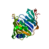 1iwlC  1iwmSC 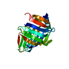 1ua8C C: citing same article ( S: Starting model for refinement |
|---|---|
| Similar structure data |


-Links
 PDBj
PDBj


- Assembly
| Deposited unit | 
| ||||||||
|---|---|---|---|---|---|---|---|---|---|
| 1 |
| ||||||||
| Unit cell |
|
-Components
| #1: Protein | Mass: 21225.721 Da / Num. of mol.: 1 / Mutation: C1A Source method: isolated from a genetically manipulated source Source: (gene. exp.) | ||||
|---|---|---|---|---|---|
| #2: Chemical |   Mass: 96.063 Da / Num. of mol.: 2 / Source method: obtained synthetically / Formula: SO4 Mass: 96.063 Da / Num. of mol.: 2 / Source method: obtained synthetically / Formula: SO4#3: Chemical | ChemComp-PG5 / | 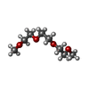  Mass: 178.226 Da / Num. of mol.: 1 / Source method: obtained synthetically / Formula: C8H18O4 Mass: 178.226 Da / Num. of mol.: 1 / Source method: obtained synthetically / Formula: C8H18O4#4: Water | ChemComp-HOH / |  Mass: 18.015 Da / Num. of mol.: 85 / Source method: isolated from a natural source / Formula: H2O Mass: 18.015 Da / Num. of mol.: 85 / Source method: isolated from a natural source / Formula: H2O |
-Experimental details
-Experiment
| Experiment | Method: X-RAY DIFFRACTION / Number of used crystals: 1 |
|---|
- Sample preparation
| Crystal | Density Matthews: 2.28 Å3/Da / Density % sol: 45.6 % | ||||||||||||||||||||||||||||||||||||||||||
|---|---|---|---|---|---|---|---|---|---|---|---|---|---|---|---|---|---|---|---|---|---|---|---|---|---|---|---|---|---|---|---|---|---|---|---|---|---|---|---|---|---|---|---|
| Crystal grow | Temperature: 293 K / Method: vapor diffusion, sitting drop / pH: 4.6 Details: PEGMME2000, sodium acetate, ammonium sulfate, sodium iodide, pH 4.6, VAPOR DIFFUSION, SITTING DROP, temperature 293K | ||||||||||||||||||||||||||||||||||||||||||
| Crystal grow | *PLUS Temperature: 20 ℃ / pH: 8 / Method: vapor diffusion, hanging drop | ||||||||||||||||||||||||||||||||||||||||||
| Components of the solutions | *PLUS
|
-Data collection
| Diffraction | Mean temperature: 90 K |
|---|---|
| Diffraction source | Source: SYNCHROTRON / Site: SPring-8  / Beamline: BL44B2 / Wavelength: 1.1 Å / Beamline: BL44B2 / Wavelength: 1.1 Å |
| Detector | Type: MARRESEARCH / Detector: CCD / Date: Jun 7, 2001 |
| Radiation | Monochromator: Si 111 / Protocol: SINGLE WAVELENGTH / Monochromatic (M) / Laue (L): M / Scattering type: x-ray |
| Radiation wavelength | Wavelength: 1.1 Å / Relative weight: 1 |
| Reflection | Resolution: 2.2→30 Å / Num. obs: 10647 / % possible obs: 98.2 % / Observed criterion σ(F): 0 / Observed criterion σ(I): 0 / Redundancy: 19.9 % / Biso Wilson estimate: 33.9 Å2 / Rsym value: 0.073 |
| Reflection shell | Resolution: 2.2→2.28 Å / Rsym value: 0.253 / % possible all: 93.4 |
| Reflection | *PLUS Highest resolution: 2.2 Å / Lowest resolution: 30 Å / Rmerge(I) obs: 0.073 |
| Reflection shell | *PLUS % possible obs: 93.4 % / Rmerge(I) obs: 0.253 |
- Processing
| Software |
| ||||||||||||||||
|---|---|---|---|---|---|---|---|---|---|---|---|---|---|---|---|---|---|
| Refinement | Method to determine structure: MOLECULAR REPLACEMENT Starting model: PDB ENTRY 1IWM (chain A) Resolution: 2.2→30 Å / Isotropic thermal model: Isotropic / Cross valid method: THROUGHOUT / σ(F): 0 / Stereochemistry target values: Engh & Huber
| ||||||||||||||||
| Displacement parameters | Biso mean: 34.4 Å2
| ||||||||||||||||
| Refine analyze |
| ||||||||||||||||
| Refinement step | Cycle: LAST / Resolution: 2.2→30 Å
| ||||||||||||||||
| Refine LS restraints |
| ||||||||||||||||
| LS refinement shell | Resolution: 2.2→2.34 Å / Rfactor Rfree error: 0.03
| ||||||||||||||||
| Refinement | *PLUS Highest resolution: 2.2 Å / Lowest resolution: 30 Å / % reflection Rfree: 5 % | ||||||||||||||||
| Solvent computation | *PLUS | ||||||||||||||||
| Displacement parameters | *PLUS | ||||||||||||||||
| LS refinement shell | *PLUS Lowest resolution: 2.28 Å |
