 Movie
Movie Controller


+ Open data

- Basic information
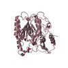
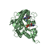
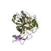
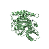
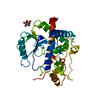
| Entry | Database: PDB / ID: 5i5h | |||||||||
|---|---|---|---|---|---|---|---|---|---|---|
| Title | Ecoli global domain 245-586 | |||||||||
 Components Components | Inner membrane protein YejM | |||||||||
 Keywords Keywords | MEMBRANE PROTEIN / Ecoli 245-586 | |||||||||
| Function / homology |  Function and homology information Function and homology informationheparan sulfate proteoglycan catabolic process / Hydrolases; Acting on ester bonds / hydrolase activity / lipid binding / plasma membrane Similarity search - Function | |||||||||
| Biological species |  | |||||||||
| Method |  X-RAY DIFFRACTION / SYNCHROTRON / MOLECULAR REPLACEMENT / Resolution: 1.65 Å X-RAY DIFFRACTION / SYNCHROTRON / MOLECULAR REPLACEMENT / Resolution: 1.65 Å | |||||||||
 Authors Authors | Dong, C. / Dong, H. | |||||||||
 Citation Citation | Journal: Sci Rep / Year: 2016 Title: Structural insights into cardiolipin transfer from the Inner membrane to the outer membrane by PbgA in Gram-negative bacteria. Authors: Dong, H. / Zhang, Z. / Tang, X. / Huang, S. / Li, H. / Peng, B. / Dong, C. | |||||||||
| History |
|
- Structure visualization
| Structure viewer | Molecule: MolmilJmol/JSmol |
|---|
- Downloads & links
-Download
| PDBx/mmCIF format | 5i5h.cif.gz | 171.5 KB | Display | PDBx/mmCIF format |
|---|---|---|---|---|
| PDB format | pdb5i5h.ent.gz | 133.2 KB | Display | PDB format |
| PDBx/mmJSON format | 5i5h.json.gz | Tree view | PDBx/mmJSON format | |
| Others |  Other downloads Other downloads |
-Validation report
| Arichive directory | https://data.pdbj.org/pub/pdb/validation_reports/i5/5i5hftp://data.pdbj.org/pub/pdb/validation_reports/i5/5i5h | HTTPS FTP |
|---|
-Related structure data
| Related structure data | 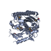 5i5dSC 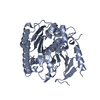 5i5fC S: Starting model for refinement C: citing same article ( |
|---|---|
| Similar structure data |


-Links
 PDBj
PDBj
- Assembly
| Deposited unit | 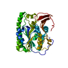
| |||||||||
|---|---|---|---|---|---|---|---|---|---|---|
| 1 |
| |||||||||
| Unit cell |
| |||||||||
| Components on special symmetry positions |
|
-Components
| #1: Protein | Mass: 67364.680 Da / Num. of mol.: 1 Source method: isolated from a genetically manipulated source Source: (gene. exp.) Strain: K12 / Gene: yejM, yejN, b2188, JW2176 / Production host: |
|---|---|
| #2: Water | ChemComp-HOH /  Mass: 18.015 Da / Num. of mol.: 495 / Source method: isolated from a natural source / Formula: H2O Mass: 18.015 Da / Num. of mol.: 495 / Source method: isolated from a natural source / Formula: H2O |
-Experimental details
-Experiment
| Experiment | Method: X-RAY DIFFRACTION / Number of used crystals: 1 |
|---|
- Sample preparation
| Crystal grow | Temperature: 310 K / Method: vapor diffusion, sitting drop / pH: 4.3 Details: 0.2 M lithium sulphate, 0.1 M sodium acetate pH 4.3 and 48% PEG400 PH range: 4.0-4.6 |
|---|
-Data collection
| Diffraction | Mean temperature: 193 K |
|---|---|
| Diffraction source | Source: SYNCHROTRON / Site: Diamond  / Beamline: I02 / Wavelength: 0.9798 Å / Beamline: I02 / Wavelength: 0.9798 Å |
| Detector | Type: DECTRIS PILATUS 6M-F / Detector: PIXEL / Date: Oct 1, 2015 |
| Radiation | Protocol: SINGLE WAVELENGTH / Monochromatic (M) / Laue (L): M / Scattering type: x-ray |
| Radiation wavelength | Wavelength: 0.9798 Å / Relative weight: 1 |
| Reflection | Resolution: 1.65→47.83 Å / % possible obs: 99.6 % / Redundancy: 9.6 % / Net I/σ(I): 17.8 |
| Reflection shell | Resolution: 1.65→1.71 Å / Redundancy: 9.6 % / % possible all: 99.2 |
- Processing
| Software |
| ||||||||||||||||||||||||||||||||||||||||||||||||||||||||||||||||||||||||||||||||||||||||||||||||||||||||||||||||||||||||||||||||||||||||||||||||||||||||||||||||||||||||||||||||||||||
|---|---|---|---|---|---|---|---|---|---|---|---|---|---|---|---|---|---|---|---|---|---|---|---|---|---|---|---|---|---|---|---|---|---|---|---|---|---|---|---|---|---|---|---|---|---|---|---|---|---|---|---|---|---|---|---|---|---|---|---|---|---|---|---|---|---|---|---|---|---|---|---|---|---|---|---|---|---|---|---|---|---|---|---|---|---|---|---|---|---|---|---|---|---|---|---|---|---|---|---|---|---|---|---|---|---|---|---|---|---|---|---|---|---|---|---|---|---|---|---|---|---|---|---|---|---|---|---|---|---|---|---|---|---|---|---|---|---|---|---|---|---|---|---|---|---|---|---|---|---|---|---|---|---|---|---|---|---|---|---|---|---|---|---|---|---|---|---|---|---|---|---|---|---|---|---|---|---|---|---|---|---|---|---|
| Refinement | Method to determine structure: MOLECULAR REPLACEMENT Starting model: 5I5D Resolution: 1.65→47.7 Å / Cor.coef. Fo:Fc: 0.973 / Cor.coef. Fo:Fc free: 0.95 / SU B: 4.04 / SU ML: 0.062 / Cross valid method: THROUGHOUT / ESU R: 0.03 / ESU R Free: 0.023 / Details: HYDROGENS HAVE BEEN ADDED IN THE RIDING POSITIONS
| ||||||||||||||||||||||||||||||||||||||||||||||||||||||||||||||||||||||||||||||||||||||||||||||||||||||||||||||||||||||||||||||||||||||||||||||||||||||||||||||||||||||||||||||||||||||
| Solvent computation | Ion probe radii: 0.8 Å / Shrinkage radii: 0.8 Å / VDW probe radii: 1.2 Å | ||||||||||||||||||||||||||||||||||||||||||||||||||||||||||||||||||||||||||||||||||||||||||||||||||||||||||||||||||||||||||||||||||||||||||||||||||||||||||||||||||||||||||||||||||||||
| Displacement parameters | Biso mean: 39.212 Å2
| ||||||||||||||||||||||||||||||||||||||||||||||||||||||||||||||||||||||||||||||||||||||||||||||||||||||||||||||||||||||||||||||||||||||||||||||||||||||||||||||||||||||||||||||||||||||
| Refinement step | Cycle: LAST / Resolution: 1.65→47.7 Å
| ||||||||||||||||||||||||||||||||||||||||||||||||||||||||||||||||||||||||||||||||||||||||||||||||||||||||||||||||||||||||||||||||||||||||||||||||||||||||||||||||||||||||||||||||||||||
| Refine LS restraints |
|