embryonic olfactory bulb interneuron precursor migration / anatomical structure arrangement / regulation of ERK5 cascade / angiotensin-activated signaling pathway involved in heart process / cerebral cortex GABAergic interneuron development / regulation of respiratory burst / positive regulation of ovarian follicle development / auditory receptor cell morphogenesis / cerebral cortex radially oriented cell migration / erythrocyte enucleation ...embryonic olfactory bulb interneuron precursor migration / anatomical structure arrangement / regulation of ERK5 cascade / angiotensin-activated signaling pathway involved in heart process / cerebral cortex GABAergic interneuron development / regulation of respiratory burst / positive regulation of ovarian follicle development / auditory receptor cell morphogenesis / cerebral cortex radially oriented cell migration / erythrocyte enucleation / regulation of neutrophil migration / negative regulation of interleukin-23 production / Activated NTRK2 signals through CDK5 / localization within membrane / kinocilium / regulation of cell adhesion involved in heart morphogenesis / interneuron migration / blue light photoreceptor activity / ruffle assembly / NTRK2 activates RAC1 / NADPH oxidase complex / cochlea morphogenesis / Inactivation of CDC42 and RAC1 / engulfment of apoptotic cell / regulation of hydrogen peroxide metabolic process / regulation of neuron maturation / respiratory burst / WNT5:FZD7-mediated leishmania damping / SEMA3A-Plexin repulsion signaling by inhibiting Integrin adhesion / cortical cytoskeleton organization / positive regulation of skeletal muscle acetylcholine-gated channel clustering / ruffle organization / epithelial cell morphogenesis / midbrain dopaminergic neuron differentiation / positive regulation of bicellular tight junction assembly / GTP-dependent protein binding / cell projection assembly / thioesterase binding / regulation of lamellipodium assembly / negative regulation of fibroblast migration / regulation of neuron migration / regulation of stress fiber assembly / RHO GTPases activate CIT / cell-cell junction organization / motor neuron axon guidance / Nef and signal transduction / PCP/CE pathway / Activation of RAC1 / hepatocyte growth factor receptor signaling pathway / sphingosine-1-phosphate receptor signaling pathway / RHO GTPases activate KTN1 / regulation of nitric oxide biosynthetic process / quinolinate biosynthetic process / DCC mediated attractive signaling / MET activates RAP1 and RAC1 / Azathioprine ADME / Sema4D mediated inhibition of cell attachment and migration / hyperosmotic response / CD28 dependent Vav1 pathway / Ephrin signaling / positive regulation of neutrophil chemotaxis / Wnt signaling pathway, planar cell polarity pathway / superoxide anion generation / regulation of receptor signaling pathway via JAK-STAT / lamellipodium assembly / NRAGE signals death through JNK / positive regulation of ruffle assembly / dendrite morphogenesis / small GTPase-mediated signal transduction / Rho GDP-dissociation inhibitor binding / Activation of RAC1 downstream of NMDARs / positive regulation of Rho protein signal transduction / positive regulation of dendritic spine development / synaptic transmission, GABAergic / pericentriolar material / positive regulation of actin filament polymerization / establishment or maintenance of cell polarity / semaphorin-plexin signaling pathway / RHO GTPases activate PAKs / Rac protein signal transduction / Sema3A PAK dependent Axon repulsion / ficolin-1-rich granule membrane / EPH-ephrin mediated repulsion of cells / regulation of postsynapse assembly / positive regulation of focal adhesion assembly / RHO GTPases Activate NADPH Oxidases / anatomical structure morphogenesis / regulation of synaptic vesicle endocytosis / regulation of neuronal synaptic plasticity / RHO GTPases Activate WASPs and WAVEs / RHO GTPases activate IQGAPs / RHO GTPases activate PKNs / PTK6 Regulates RHO GTPases, RAS GTPase and MAP kinases / GPVI-mediated activation cascade / phagocytic cup / positive regulation of lamellipodium assembly / positive regulation of stress fiber assembly / cell projection / actin filament polymerization / RAC1 GTPase cycle Similarity search - Function
PAS domain / PAS-associated, C-terminal / PAC domain profile. / Small GTPase Rho / Small GTPase Rho domain profile. / PAC motif / Motif C-terminal to PAS motifs (likely to contribute to PAS structural domain) / PAS domain / Beta-Lactamase / PAS domain ...PAS domain / PAS-associated, C-terminal / PAC domain profile. / Small GTPase Rho / Small GTPase Rho domain profile. / PAC motif / Motif C-terminal to PAS motifs (likely to contribute to PAS structural domain) / PAS domain / Beta-Lactamase / PAS domain / PAS repeat profile. / PAS domain / PAS domain superfamily / Rho (Ras homology) subfamily of Ras-like small GTPases / Ras subfamily of RAS small GTPases / Small GTPase / Ras family / Rab subfamily of small GTPases / Small GTP-binding protein domain / P-loop containing nucleotide triphosphate hydrolases / Serine/threonine-protein kinase, active site / Serine/Threonine protein kinases active-site signature. / Protein kinase domain / Serine/Threonine protein kinases, catalytic domain / Protein kinase, ATP binding site / Protein kinases ATP-binding region signature. / Protein kinase domain profile. / Protein kinase domain / Protein kinase-like domain superfamily / Rossmann fold / P-loop containing nucleoside triphosphate hydrolase / 2-Layer Sandwich / 3-Layer(aba) Sandwich / Alpha Beta Similarity search - Domain/homology
In the structure databanks used in Yorodumi, some data are registered as the other names, "COVID-19 virus" and "2019-nCoV". Here are the details of the virus and the list of structure data.
Jan 31, 2019. EMDB accession codes are about to change! (news from PDBe EMDB page)
EMDB accession codes are about to change! (news from PDBe EMDB page)
The allocation of 4 digits for EMDB accession codes will soon come to an end. Whilst these codes will remain in use, new EMDB accession codes will include an additional digit and will expand incrementally as the available range of codes is exhausted. The current 4-digit format prefixed with “EMD-” (i.e. EMD-XXXX) will advance to a 5-digit format (i.e. EMD-XXXXX), and so on. It is currently estimated that the 4-digit codes will be depleted around Spring 2019, at which point the 5-digit format will come into force.
The EM Navigator/Yorodumi systems omit the EMD- prefix.
Related info.:Q: What is EMD? / ID/Accession-code notation in Yorodumi/EM Navigator
Yorodumi is a browser for structure data from EMDB, PDB, SASBDB, etc.
This page is also the successor to EM Navigator detail page, and also detail information page/front-end page for Omokage search.
The word "yorodu" (or yorozu) is an old Japanese word meaning "ten thousand". "mi" (miru) is to see.
Related info.:EMDB / PDB / SASBDB / Comparison of 3 databanks / Yorodumi Search / Aug 31, 2016. New EM Navigator & Yorodumi / Yorodumi Papers / Jmol/JSmol / Function and homology information / Changes in new EM Navigator and Yorodumi
 Movie
Movie Controller

 Yorodumi
 Open data
 Basic information
 Components
 Keywords
 Function and homology information
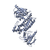
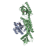
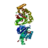
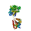
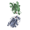
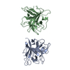
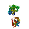
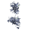
 Homo sapiens (human)
 X-RAY DIFFRACTION /
  Authors
 Citation
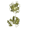
 Structure visualization
 Downloads & links
 Other downloads
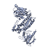


 PDBj
PDBj


 Assembly


 Mass: 523.180 Da / Num. of mol.: 1 / Source method: obtained synthetically / Formula: C10H16N5O14P3 / Comment: GTP, energy-carrying molecule*YM
 Mass: 456.344 Da / Num. of mol.: 1 / Source method: obtained synthetically / Formula: C17H21N4O9P
 Mass: 24.305 Da / Num. of mol.: 1 / Source method: obtained synthetically / Formula: Mg
 Mass: 40.078 Da / Num. of mol.: 2 / Source method: obtained synthetically / Formula: Ca
 Sample preparation
 / Beamline: X10SA / Wavelength: 0.97885 Å
 Processing