 Movie
Movie Controller

[English] 日本語
 Yorodumi
- PDB-3csq: Crystal and cryoEM structural studies of a cell wall degrading en... -
+ Open data

- Basic information
| Entry | Database: PDB / ID: 3csq | ||||||
|---|---|---|---|---|---|---|---|
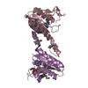
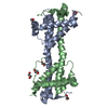
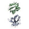
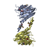
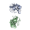
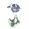
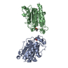
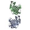
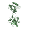
| Title | Crystal and cryoEM structural studies of a cell wall degrading enzyme in the bacteriophage phi29 tail | ||||||
 Components Components | Morphogenesis protein 1 | ||||||
 Keywords Keywords | HYDROLASE / infection / phi29 / Late protein | ||||||
| Function / homology |  Function and homology information Function and homology informationvirus tail, tip / virus tail fiber assembly / symbiont entry into host cell via disruption of host cell wall peptidoglycan / symbiont genome ejection through host cell envelope, short tail mechanism / symbiont entry into host cell via disruption of host cell envelope / hydrolase activity, acting on glycosyl bonds / Hydrolases; Glycosylases; Glycosidases, i.e. enzymes that hydrolyse O- and S-glycosyl compounds / Hydrolases; Acting on peptide bonds (peptidases) / cell wall organization / metallopeptidase activity ...virus tail, tip / virus tail fiber assembly / symbiont entry into host cell via disruption of host cell wall peptidoglycan / symbiont genome ejection through host cell envelope, short tail mechanism / symbiont entry into host cell via disruption of host cell envelope / hydrolase activity, acting on glycosyl bonds / Hydrolases; Glycosylases; Glycosidases, i.e. enzymes that hydrolyse O- and S-glycosyl compounds / Hydrolases; Acting on peptide bonds (peptidases) / cell wall organization / metallopeptidase activity / killing of cells of another organism / defense response to bacterium / proteolysis / metal ion binding Similarity search - Function | ||||||
| Biological species |  Bacteriophage phi-29 (virus) Bacteriophage phi-29 (virus) | ||||||
| Method |  X-RAY DIFFRACTION / SYNCHROTRON / SIR, SAD / Resolution: 1.8 Å X-RAY DIFFRACTION / SYNCHROTRON / SIR, SAD / Resolution: 1.8 Å | ||||||
 Authors Authors | Xiang, Y. / Rossmann, M.G. | ||||||
 Citation Citation | Journal: Proc Natl Acad Sci U S A / Year: 2008 Title: Crystal and cryoEM structural studies of a cell wall degrading enzyme in the bacteriophage phi29 tail. Authors: Ye Xiang / Marc C Morais / Daniel N Cohen / Valorie D Bowman / Dwight L Anderson / Michael G Rossmann /  Abstract: The small bacteriophage phi29 must penetrate the approximately 250-A thick external peptidoglycan cell wall and cell membrane of the Gram-positive Bacillus subtilis, before ejecting its dsDNA genome ...The small bacteriophage phi29 must penetrate the approximately 250-A thick external peptidoglycan cell wall and cell membrane of the Gram-positive Bacillus subtilis, before ejecting its dsDNA genome through its tail into the bacterial cytoplasm. The tail of bacteriophage phi29 is noncontractile and approximately 380 A long. A 1.8-A resolution crystal structure of gene product 13 (gp13) shows that this tail protein has spatially well separated N- and C-terminal domains, whose structures resemble lysozyme-like enzymes and metallo-endopeptidases, respectively. CryoEM reconstructions of the WT bacteriophage and mutant bacteriophages missing some or most of gp13 shows that this enzyme is located at the distal end of the phi29 tail knob. This finding suggests that gp13 functions as a tail-associated, peptidoglycan-degrading enzyme able to cleave both the polysaccharide backbone and peptide cross-links of the peptidoglycan cell wall. Comparisons of the gp13(-) mutants with the phi29 mature and emptied phage structures suggest the sequence of events that occur during the penetration of the tail through the peptidoglycan layer. | ||||||
| History |
|
- Structure visualization
| Structure viewer | Molecule: MolmilJmol/JSmol |
|---|
- Downloads & links
-Download
| PDBx/mmCIF format | 3csq.cif.gz | 263.4 KB | Display | PDBx/mmCIF format |
|---|---|---|---|---|
| PDB format | pdb3csq.ent.gz | 214.4 KB | Display | PDB format |
| PDBx/mmJSON format | 3csq.json.gz | Tree view | PDBx/mmJSON format | |
| Others |  Other downloads Other downloads |
-Validation report
| Arichive directory | https://data.pdbj.org/pub/pdb/validation_reports/cs/3csqftp://data.pdbj.org/pub/pdb/validation_reports/cs/3csq | HTTPS FTP |
|---|
-Related structure data
| Related structure data | 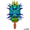 1506C 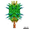 5010C 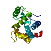 3csrC 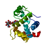 3cszC 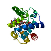 3ct0C 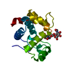 3ct1C 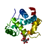 3ct5C C: citing same article ( |
|---|---|
| Similar structure data |
-Links
 PDBj
PDBj- Assembly
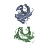
| Deposited unit | 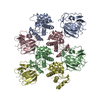
| ||||||||
|---|---|---|---|---|---|---|---|---|---|
| 1 | 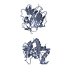
| ||||||||
| 2 | 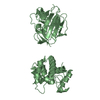
| ||||||||
| 3 | 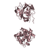
| ||||||||
| 4 | 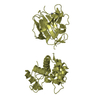
| ||||||||
| Unit cell |
|
-Components
| #1: Protein | Mass: 37648.074 Da / Num. of mol.: 4 Source method: isolated from a genetically manipulated source Source: (gene. exp.) Bacteriophage phi-29 (virus) / Gene: 13 / Plasmid: PTYB1 / Production host:  #2: Chemical | ChemComp-ZN /   Mass: 65.409 Da / Num. of mol.: 4 / Source method: obtained synthetically / Formula: Zn Mass: 65.409 Da / Num. of mol.: 4 / Source method: obtained synthetically / Formula: Zn#3: Water | ChemComp-HOH / |  Mass: 18.015 Da / Num. of mol.: 216 / Source method: isolated from a natural source / Formula: H2O Mass: 18.015 Da / Num. of mol.: 216 / Source method: isolated from a natural source / Formula: H2OSequence details | AUTORS STATE THAT RESIDUE 89 SHOULD BE ASN ACCORDING TO THEIR SEQUENCING | |
|---|
-Experimental details
-Experiment
| Experiment | Method: X-RAY DIFFRACTION / Number of used crystals: 1 |
|---|
- Sample preparation
| Crystal | Density Matthews: 2.05 Å3/Da / Density % sol: 40.09 % |
|---|---|
| Crystal grow | Temperature: 293 K / Method: vapor diffusion, sitting drop / pH: 8 Details: 100mM Tris-HCl, 200mM MgCl2, 28% (w/v) PEG4000, pH 8.0, VAPOR DIFFUSION, SITTING DROP, temperature 293K |
-Data collection
| Diffraction | Mean temperature: 100 K |
|---|---|
| Diffraction source | Source: SYNCHROTRON / Site: APS / Beamline: 23-ID-D / Wavelength: 1.03 Å |
| Detector | Type: MARMOSAIC 300 mm CCD / Detector: CCD / Date: Jun 21, 2007 / Details: mirrors |
| Radiation | Monochromator: Si 111 CHANNEL / Protocol: SINGLE WAVELENGTH / Monochromatic (M) / Laue (L): M / Scattering type: x-ray |
| Radiation wavelength | Wavelength: 1.03 Å / Relative weight: 1 |
| Reflection | Resolution: 1.8→19.9 Å / Num. obs: 109340 / Rmerge(I) obs: 0.055 / Net I/σ(I): 20 |
| Reflection shell | Resolution: 1.8→1.85 Å / Rmerge(I) obs: 0.109 / Mean I/σ(I) obs: 9.7 |
- Processing
| Software |
| ||||||||||||||||||||||||||||||||||||||||||||||||||||||||||||||||||||||||||||||||||||||||||||||||||||||||||||||||||||||||||||||||||||||||||||||||||||||||||||||||||||||||||
|---|---|---|---|---|---|---|---|---|---|---|---|---|---|---|---|---|---|---|---|---|---|---|---|---|---|---|---|---|---|---|---|---|---|---|---|---|---|---|---|---|---|---|---|---|---|---|---|---|---|---|---|---|---|---|---|---|---|---|---|---|---|---|---|---|---|---|---|---|---|---|---|---|---|---|---|---|---|---|---|---|---|---|---|---|---|---|---|---|---|---|---|---|---|---|---|---|---|---|---|---|---|---|---|---|---|---|---|---|---|---|---|---|---|---|---|---|---|---|---|---|---|---|---|---|---|---|---|---|---|---|---|---|---|---|---|---|---|---|---|---|---|---|---|---|---|---|---|---|---|---|---|---|---|---|---|---|---|---|---|---|---|---|---|---|---|---|---|---|---|---|---|
| Refinement | Method to determine structure: SIR, SAD / Resolution: 1.8→19.9 Å / Cor.coef. Fo:Fc: 0.902 / Cor.coef. Fo:Fc free: 0.863 / SU B: 3.825 / SU ML: 0.122 / Cross valid method: THROUGHOUT / ESU R: 0.184 / ESU R Free: 0.167 / Stereochemistry target values: MAXIMUM LIKELIHOOD
| ||||||||||||||||||||||||||||||||||||||||||||||||||||||||||||||||||||||||||||||||||||||||||||||||||||||||||||||||||||||||||||||||||||||||||||||||||||||||||||||||||||||||||
| Solvent computation | Ion probe radii: 0.8 Å / Shrinkage radii: 0.8 Å / VDW probe radii: 1.2 Å / Solvent model: BABINET MODEL WITH MASK | ||||||||||||||||||||||||||||||||||||||||||||||||||||||||||||||||||||||||||||||||||||||||||||||||||||||||||||||||||||||||||||||||||||||||||||||||||||||||||||||||||||||||||
| Displacement parameters | Biso mean: 18.084 Å2
| ||||||||||||||||||||||||||||||||||||||||||||||||||||||||||||||||||||||||||||||||||||||||||||||||||||||||||||||||||||||||||||||||||||||||||||||||||||||||||||||||||||||||||
| Refinement step | Cycle: LAST / Resolution: 1.8→19.9 Å
| ||||||||||||||||||||||||||||||||||||||||||||||||||||||||||||||||||||||||||||||||||||||||||||||||||||||||||||||||||||||||||||||||||||||||||||||||||||||||||||||||||||||||||
| Refine LS restraints |
| ||||||||||||||||||||||||||||||||||||||||||||||||||||||||||||||||||||||||||||||||||||||||||||||||||||||||||||||||||||||||||||||||||||||||||||||||||||||||||||||||||||||||||
| LS refinement shell | Resolution: 1.8→1.846 Å / Total num. of bins used: 20
|
