 Movie
Movie Controller
 Structure viewers
 About Yorodumi Papers


+Search query
-Structure paper
| Title | Crystal and cryoEM structural studies of a cell wall degrading enzyme in the bacteriophage phi29 tail. |
|---|---|
| Journal, issue, pages | Proc Natl Acad Sci U S A, Vol. 105, Issue 28, Page 9552-9557, Year 2008 |
| Publish date | Jul 15, 2008 |
 Authors Authors | Ye Xiang / Marc C Morais / Daniel N Cohen / Valorie D Bowman / Dwight L Anderson / Michael G Rossmann /  |
| PubMed Abstract | The small bacteriophage phi29 must penetrate the approximately 250-A thick external peptidoglycan cell wall and cell membrane of the Gram-positive Bacillus subtilis, before ejecting its dsDNA genome ...The small bacteriophage phi29 must penetrate the approximately 250-A thick external peptidoglycan cell wall and cell membrane of the Gram-positive Bacillus subtilis, before ejecting its dsDNA genome through its tail into the bacterial cytoplasm. The tail of bacteriophage phi29 is noncontractile and approximately 380 A long. A 1.8-A resolution crystal structure of gene product 13 (gp13) shows that this tail protein has spatially well separated N- and C-terminal domains, whose structures resemble lysozyme-like enzymes and metallo-endopeptidases, respectively. CryoEM reconstructions of the WT bacteriophage and mutant bacteriophages missing some or most of gp13 shows that this enzyme is located at the distal end of the phi29 tail knob. This finding suggests that gp13 functions as a tail-associated, peptidoglycan-degrading enzyme able to cleave both the polysaccharide backbone and peptide cross-links of the peptidoglycan cell wall. Comparisons of the gp13(-) mutants with the phi29 mature and emptied phage structures suggest the sequence of events that occur during the penetration of the tail through the peptidoglycan layer. |
 External links External links | Proc Natl Acad Sci U S A / PubMed:18606992 / PubMed Central |
| Methods | EM (single particle) / X-ray diffraction |
| Resolution | 1.37 - 35.0 Å |
| Structure data | 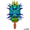 EMDB-1506: 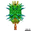 EMDB-5010: 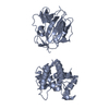 PDB-3csq: 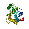 PDB-3csr: 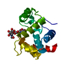 PDB-3csz: 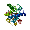 PDB-3ct0: 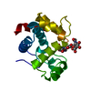 PDB-3ct1: 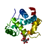 PDB-3ct5: |
| Chemicals |  ChemComp-ZN:  ChemComp-HOH: |
| Source |
|
 Keywords Keywords | HYDROLASE / infection / phi29 / Late protein / cell wall |
 bacteriophage phi-29 (virus)