 Movie
Movie Controller

[English] 日本語
 Yorodumi
- EMDB-1506: Crystal and cryoEM structural studies of a cell wall degrading en... -
+ Open data

- Basic information
| Entry | Database: EMDB / ID: EMD-1506 | |||||||||
|---|---|---|---|---|---|---|---|---|---|---|
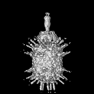
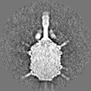
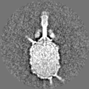
| Title | Crystal and cryoEM structural studies of a cell wall degrading enzyme in the bacteriophage phi29 tail | |||||||||
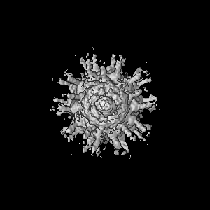
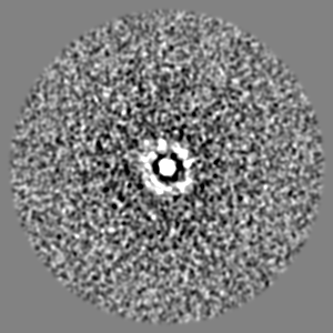
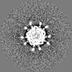
 Map data Map data | This is an image of a surface rendered top-view of bacteriophage phi29 mutant sus13(330) | |||||||||
 Sample Sample |
| |||||||||
 Keywords Keywords | Cell wall / phi29 / hydrolase / infection / structure | |||||||||
| Biological species |   Bacillus phage phi29 (virus) Bacillus phage phi29 (virus) | |||||||||
| Method | single particle reconstruction / cryo EM / Resolution: 30.0 Å | |||||||||
 Authors Authors | Xiang Y / Morais MC / Cohen DN / Bowman VD / Anderson DL / Rossmann MG | |||||||||
 Citation Citation | Journal: Proc Natl Acad Sci U S A / Year: 2008 Title: Crystal and cryoEM structural studies of a cell wall degrading enzyme in the bacteriophage phi29 tail. Authors: Ye Xiang / Marc C Morais / Daniel N Cohen / Valorie D Bowman / Dwight L Anderson / Michael G Rossmann /  Abstract: The small bacteriophage phi29 must penetrate the approximately 250-A thick external peptidoglycan cell wall and cell membrane of the Gram-positive Bacillus subtilis, before ejecting its dsDNA genome ...The small bacteriophage phi29 must penetrate the approximately 250-A thick external peptidoglycan cell wall and cell membrane of the Gram-positive Bacillus subtilis, before ejecting its dsDNA genome through its tail into the bacterial cytoplasm. The tail of bacteriophage phi29 is noncontractile and approximately 380 A long. A 1.8-A resolution crystal structure of gene product 13 (gp13) shows that this tail protein has spatially well separated N- and C-terminal domains, whose structures resemble lysozyme-like enzymes and metallo-endopeptidases, respectively. CryoEM reconstructions of the WT bacteriophage and mutant bacteriophages missing some or most of gp13 shows that this enzyme is located at the distal end of the phi29 tail knob. This finding suggests that gp13 functions as a tail-associated, peptidoglycan-degrading enzyme able to cleave both the polysaccharide backbone and peptide cross-links of the peptidoglycan cell wall. Comparisons of the gp13(-) mutants with the phi29 mature and emptied phage structures suggest the sequence of events that occur during the penetration of the tail through the peptidoglycan layer. | |||||||||
| History |
|
- Structure visualization
| Movie |
 Movie viewer Movie viewer |
|---|---|
| Structure viewer | EM map: SurfViewMolmilJmol/JSmol |
| Supplemental images |

- Downloads & links
-EMDB archive
| Map data | emd_1506.map.gz | 76.9 MB | EMDB map data format | |
|---|---|---|---|---|
| Header (meta data) | emd-1506-v30.xmlemd-1506.xml | 9.7 KB 9.7 KB | Display Display | EMDB header |
| Images | 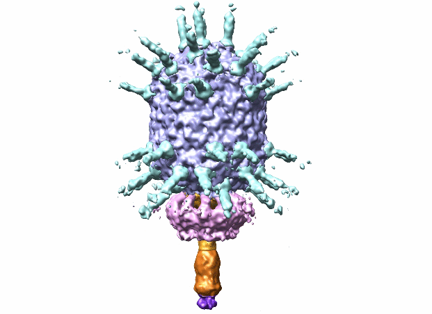 1506.gif 1506.gif | 53.1 KB | ||
| Archive directory |  http://ftp.pdbj.org/pub/emdb/structures/EMD-1506ftp://ftp.pdbj.org/pub/emdb/structures/EMD-1506 http://ftp.pdbj.org/pub/emdb/structures/EMD-1506ftp://ftp.pdbj.org/pub/emdb/structures/EMD-1506 | HTTPS FTP |
-Related structure data
| Related structure data | 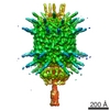 5010C  3csqC  3csrC 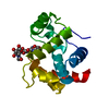 3cszC 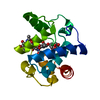 3ct0C 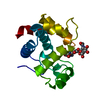 3ct1C 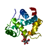 3ct5C C: citing same article ( |
|---|---|
| Similar structure data |
-Links
| EMDB pages | EMDB (EBI/PDBe) / EMDataResource |
|---|
-Map
| File | Download / File: emd_1506.map.gz / Format: CCP4 / Size: 100.6 MB / Type: IMAGE STORED AS FLOATING POINT NUMBER (4 BYTES) | ||||||||||||||||||||||||||||||||||||||||||||||||||||||||||||||||||||
|---|---|---|---|---|---|---|---|---|---|---|---|---|---|---|---|---|---|---|---|---|---|---|---|---|---|---|---|---|---|---|---|---|---|---|---|---|---|---|---|---|---|---|---|---|---|---|---|---|---|---|---|---|---|---|---|---|---|---|---|---|---|---|---|---|---|---|---|---|---|
| Annotation | This is an image of a surface rendered top-view of bacteriophage phi29 mutant sus13(330) | ||||||||||||||||||||||||||||||||||||||||||||||||||||||||||||||||||||
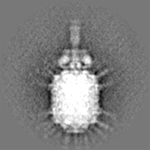
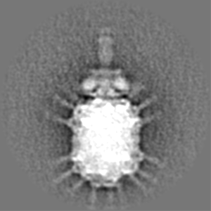
| Projections & slices | Image control
Images are generated by Spider. | ||||||||||||||||||||||||||||||||||||||||||||||||||||||||||||||||||||
| Voxel size | X=Y=Z: 4.24 Å | ||||||||||||||||||||||||||||||||||||||||||||||||||||||||||||||||||||
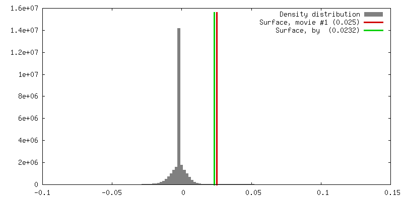
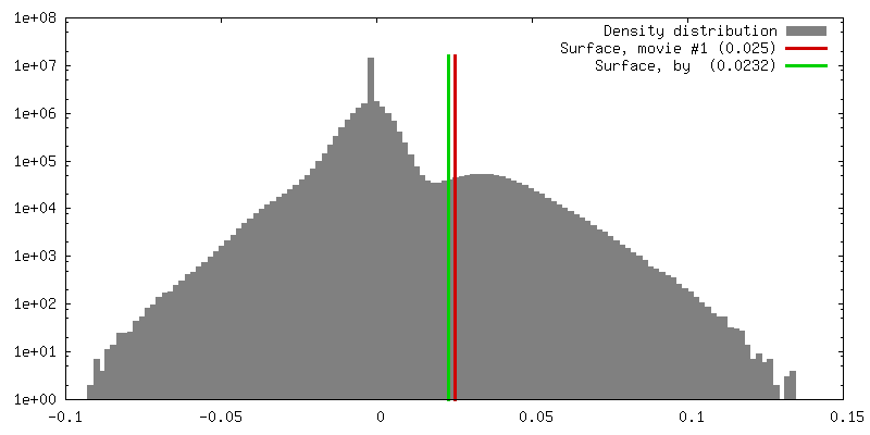
| Density |
| ||||||||||||||||||||||||||||||||||||||||||||||||||||||||||||||||||||
| Symmetry | Space group: 1 | ||||||||||||||||||||||||||||||||||||||||||||||||||||||||||||||||||||
| Details | EMDB XML:
CCP4 map header:
| ||||||||||||||||||||||||||||||||||||||||||||||||||||||||||||||||||||
 Z (Sec.)
 Y (Row.)
 X (Col.)


-Supplemental data
- Sample components
-Entire : Bacteriophage phi29 mutant sus13(330)
| Entire | Name: Bacteriophage phi29 mutant sus13(330) |
|---|---|
| Components |
|
-Supramolecule #1000: Bacteriophage phi29 mutant sus13(330)
| Supramolecule | Name: Bacteriophage phi29 mutant sus13(330) / type: sample / ID: 1000 Oligomeric state: capsid protein forms t3 q5 prolate icosahedron Number unique components: 9 |
|---|---|
| Molecular weight | Theoretical: 35.2 MDa |
-Supramolecule #1: Bacillus phage phi29
| Supramolecule | Name: Bacillus phage phi29 / type: virus / ID: 1 Details: Fibered bacteriophage phi29 mutant sus13(330). A large fragment of gene product 13 could be detected in the mutant particles. NCBI-ID: 10756 / Sci species name: Bacillus phage phi29 / Virus type: VIRUS-LIKE PARTICLE / Virus isolate: SPECIES / Virus enveloped: No / Virus empty: No |
|---|---|
| Host (natural) | Organism:  |
| Molecular weight | Theoretical: 35.2 MDa |
| Virus shell | Shell ID: 1 / Name: Phi29 sus13330 / Diameter: 530 Å / T number (triangulation number): 3 |
-Experimental details
-Structure determination
| Method | cryo EM |
|---|---|
 Processing Processing | single particle reconstruction |
| Aggregation state | particle |
-Sample preparation
| Concentration | 1 mg/mL |
|---|---|
| Buffer | pH: 7.8 / Details: 25mM Tris-HCl pH 7.8, 50mM NaCl, 5mM MgCl2 |
| Grid | Details: holey carbon |
| Vitrification | Cryogen name: ETHANE / Chamber temperature: 113 K / Instrument: OTHER |
- Electron microscopy
| Microscope | FEI/PHILIPS CM300FEG/T |
|---|---|
| Image recording | Digitization - Scanner: ZEISS SCAI / Digitization - Sampling interval: 7 µm / Number real images: 70 / Average electron dose: 20 e/Å2 / Details: after scanning, images binned by a factor of 2 / Bits/pixel: 8 |
| Electron beam | Acceleration voltage: 300 kV / Electron source:  FIELD EMISSION GUN FIELD EMISSION GUN |
| Electron optics | Illumination mode: FLOOD BEAM / Imaging mode: BRIGHT FIELD / Nominal defocus max: 3.3 µm / Nominal defocus min: 1.5 µm |
| Sample stage | Specimen holder: Side entry liquid nitrogen-cooled cryo specimen holder Specimen holder model: GATAN LIQUID NITROGEN |
-Image processing
| CTF correction | Details: phase flip |
|---|---|
| Final reconstruction | Applied symmetry - Point group: C1 (asymmetric) / Algorithm: OTHER / Resolution.type: BY AUTHOR / Resolution: 30.0 Å / Resolution method: FSC 0.5 CUT-OFF / Software - Name: EMAN / Details: asymmetric / Number images used: 5188 |
| Final two d classification | Number classes: 250 |
-Atomic model buiding 1
| Initial model | PDB ID: Chain - Chain ID: A |
|---|---|
| Software | Name: emfit |
| Details | PDBEntryID_givenInChain. Protocol: Rigid Body. Two possible biological functional units were fitted into the em map |
| Refinement | Space: REAL / Protocol: RIGID BODY FIT / Target criteria: Density distribution |
