 Movie
Movie Controller


+ Open data

- Basic information
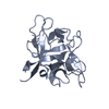
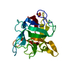
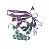
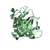
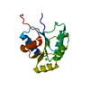
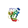
| Entry | Database: PDB / ID: 2bea | ||||||
|---|---|---|---|---|---|---|---|
| Title | Crystal structure of Asn14 to Gly mutant of WCI | ||||||
 Components Components | Chymotrypsin inhibitor 3 | ||||||
 Keywords Keywords | HYDROLASE INHIBITOR / Beta trefoil / spacer / mutant | ||||||
| Function / homology |  Function and homology information Function and homology information | ||||||
| Biological species |   Psophocarpus tetragonolobus (winged bean) Psophocarpus tetragonolobus (winged bean) | ||||||
| Method |  X-RAY DIFFRACTION / MOLECULAR REPLACEMENT / Resolution: 2.35 Å X-RAY DIFFRACTION / MOLECULAR REPLACEMENT / Resolution: 2.35 Å | ||||||
 Authors Authors | Dattagupta, J.K. / Sen, U. / Dasgupta, J. / Khamrui, S. | ||||||
 Citation Citation | Journal: Biochemistry / Year: 2006 Title: Spacer Asn Determines the Fate of Kunitz (STI) Inhibitors, as Revealed by Structural and Biochemical Studies on WCI Mutants. Authors: Dasgupta, J. / Khamrui, S. / Dattagupta, J.K. / Sen, U. #1: Journal: Biochim.Biophys.Acta / Year: 2005Title: Single mutation at P1 of a chymotrypsin inhibitor changes it to a trypsin inhibitor: X-ray structural (2.15 A) and biochemical basis. Authors: Khamrui, S. / Dasgupta, J. / Dattagupta, J.K. / Sen, U. | ||||||
| History |
|
- Structure visualization
| Structure viewer | Molecule: MolmilJmol/JSmol |
|---|
- Downloads & links
-Download
| PDBx/mmCIF format | 2bea.cif.gz | 77.4 KB | Display | PDBx/mmCIF format |
|---|---|---|---|---|
| PDB format | pdb2bea.ent.gz | 58.3 KB | Display | PDB format |
| PDBx/mmJSON format | 2bea.json.gz | Tree view | PDBx/mmJSON format | |
| Others |  Other downloads Other downloads |
-Validation report
| Arichive directory | https://data.pdbj.org/pub/pdb/validation_reports/be/2beaftp://data.pdbj.org/pub/pdb/validation_reports/be/2bea | HTTPS FTP |
|---|
-Related structure data


-Links
 PDBj
PDBj- Assembly
| Deposited unit | 
| ||||||||
|---|---|---|---|---|---|---|---|---|---|
| 1 | 
| ||||||||
| 2 | 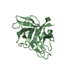
| ||||||||
| Unit cell |
|
-Components
| #1: Protein | Mass: 20617.279 Da / Num. of mol.: 2 / Mutation: N14G Source method: isolated from a genetically manipulated source Source: (gene. exp.) Psophocarpus tetragonolobus (winged bean)Species (production host): Escherichia coli / Production host:  #2: Water | ChemComp-HOH / |  Mass: 18.015 Da / Num. of mol.: 282 / Source method: isolated from a natural source / Formula: H2O Mass: 18.015 Da / Num. of mol.: 282 / Source method: isolated from a natural source / Formula: H2OHas protein modification | Y | |
|---|
-Experimental details
-Experiment
| Experiment | Method: X-RAY DIFFRACTION / Number of used crystals: 1 |
|---|
- Sample preparation
| Crystal | Density Matthews: 1.976368 Å3/Da / Density % sol: 37.764629 % |
|---|
-Data collection
| Diffraction | Mean temperature: 100 K |
|---|---|
| Diffraction source | Source: ROTATING ANODE / Type: RIGAKU RU200 / Wavelength: 1.54 Å |
| Detector | Type: MARRESEARCH / Detector: IMAGE PLATE / Date: Jan 10, 2005 |
| Radiation | Protocol: SINGLE WAVELENGTH / Monochromatic (M) / Laue (L): M / Scattering type: x-ray |
| Radiation wavelength | Wavelength: 1.54 Å / Relative weight: 1 |
| Reflection | Resolution: 2.35→15 Å / Num. all: 12855 / Num. obs: 11423 / % possible obs: 88.9 % / Observed criterion σ(F): 0 / Observed criterion σ(I): 0 / Biso Wilson estimate: 40.8 Å2 / Rmerge(I) obs: 0.044 |
- Processing
| Software |
| ||||||||||||||||||||||||||||||||||||||||||||||||||||||||||||||||||||||||||||||||
|---|---|---|---|---|---|---|---|---|---|---|---|---|---|---|---|---|---|---|---|---|---|---|---|---|---|---|---|---|---|---|---|---|---|---|---|---|---|---|---|---|---|---|---|---|---|---|---|---|---|---|---|---|---|---|---|---|---|---|---|---|---|---|---|---|---|---|---|---|---|---|---|---|---|---|---|---|---|---|---|---|---|
| Refinement | Method to determine structure: MOLECULAR REPLACEMENT / Resolution: 2.35→14.77 Å / Rfactor Rfree error: 0.011 / Data cutoff high absF: 752257 / Data cutoff low absF: 0 / Isotropic thermal model: RESTRAINED / Cross valid method: THROUGHOUT / σ(F): 0 / Stereochemistry target values: Engh & Huber
| ||||||||||||||||||||||||||||||||||||||||||||||||||||||||||||||||||||||||||||||||
| Solvent computation | Solvent model: FLAT MODEL / Bsol: 52.2231 Å2 / ksol: 0.281937 e/Å3 | ||||||||||||||||||||||||||||||||||||||||||||||||||||||||||||||||||||||||||||||||
| Displacement parameters | Biso mean: 46.4 Å2
| ||||||||||||||||||||||||||||||||||||||||||||||||||||||||||||||||||||||||||||||||
| Refine analyze |
| ||||||||||||||||||||||||||||||||||||||||||||||||||||||||||||||||||||||||||||||||
| Refinement step | Cycle: LAST / Resolution: 2.35→14.77 Å
| ||||||||||||||||||||||||||||||||||||||||||||||||||||||||||||||||||||||||||||||||
| Refine LS restraints |
| ||||||||||||||||||||||||||||||||||||||||||||||||||||||||||||||||||||||||||||||||
| LS refinement shell | Resolution: 2.35→2.5 Å / Rfactor Rfree error: 0.049 / Total num. of bins used: 6
| ||||||||||||||||||||||||||||||||||||||||||||||||||||||||||||||||||||||||||||||||
| Xplor file |
|
