 Movie
Movie Controller


+ Open data

- Basic information
| Entry | Database: PDB / ID: 3gaq | ||||||
|---|---|---|---|---|---|---|---|
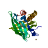
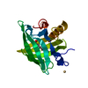
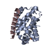
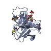
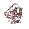
| Title | Female-specific Histamine-Binding Protein, D24R Mutant | ||||||
 Components Components | Female-specific histamine-binding protein 2 | ||||||
 Keywords Keywords | IMMUNE SYSTEM / Lipocalin / Beta barrel | ||||||
| Function / homology |  Function and homology information Function and homology informationamine binding / symbiont-mediated perturbation of host defenses / extracellular region Similarity search - Function | ||||||
| Biological species |  Rhipicephalus appendiculatus (arthropod) Rhipicephalus appendiculatus (arthropod) | ||||||
| Method |  X-RAY DIFFRACTION / MOLECULAR REPLACEMENT / molecular replacement / Resolution: 2.25 Å X-RAY DIFFRACTION / MOLECULAR REPLACEMENT / molecular replacement / Resolution: 2.25 Å | ||||||
 Authors Authors | Dennis, C.A. / Homans, S.W. | ||||||
 Citation Citation | Journal: To be Published Title: Entropic contributions to binding in a 'Hydrophilic' Ligand-Protein Interaction Authors: Syme, N.R. / Dennis, C.A. / Bronowska, A. / Paesen, G. / Homans, S.W. | ||||||
| History |
|
- Structure visualization
| Structure viewer | Molecule: MolmilJmol/JSmol |
|---|
- Downloads & links
-Download
| PDBx/mmCIF format | 3gaq.cif.gz | 83.7 KB | Display | PDBx/mmCIF format |
|---|---|---|---|---|
| PDB format | pdb3gaq.ent.gz | 63.2 KB | Display | PDB format |
| PDBx/mmJSON format | 3gaq.json.gz | Tree view | PDBx/mmJSON format | |
| Others |  Other downloads Other downloads |
-Validation report
| Arichive directory | https://data.pdbj.org/pub/pdb/validation_reports/ga/3gaqftp://data.pdbj.org/pub/pdb/validation_reports/ga/3gaq | HTTPS FTP |
|---|
-Related structure data
| Related structure data |  3g7xC  1qftS C: citing same article ( S: Starting model for refinement |
|---|---|
| Similar structure data |


-Links
 PDBj
PDBj

- Assembly
| Deposited unit | 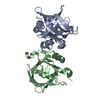
| ||||||||
|---|---|---|---|---|---|---|---|---|---|
| 1 | 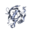
| ||||||||
| 2 | 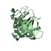
| ||||||||
| Unit cell |
|
-Components
| #1: Protein | Mass: 19597.230 Da / Num. of mol.: 2 / Mutation: D24R Source method: isolated from a genetically manipulated source Source: (gene. exp.) Rhipicephalus appendiculatus (arthropod)Plasmid: pET23a / Production host:  #2: Water | ChemComp-HOH / |  Mass: 18.015 Da / Num. of mol.: 201 / Source method: isolated from a natural source / Formula: H2O Mass: 18.015 Da / Num. of mol.: 201 / Source method: isolated from a natural source / Formula: H2OHas protein modification | Y | |
|---|
-Experimental details
-Experiment
| Experiment | Method: X-RAY DIFFRACTION / Number of used crystals: 1 |
|---|
- Sample preparation
| Crystal | Density Matthews: 2.55 Å3/Da / Density % sol: 51.82 % |
|---|---|
| Crystal grow | Temperature: 290 K / Method: vapor diffusion, hanging drop Details: Protein at 10 mg/ml mixed in a 1:1 ratio with reservoir containing: 2.6 M Ammonium Sulphate, 0.1 M Bicine, pH 8.2, and 10 % isopropanol, VAPOR DIFFUSION, HANGING DROP, temperature 290K |
-Data collection
| Diffraction | Mean temperature: 100 K |
|---|---|
| Diffraction source | Source: ROTATING ANODE / Type: RIGAKU RUH3R / Wavelength: 1.54 Å |
| Detector | Type: RIGAKU RAXIS IV++ / Detector: IMAGE PLATE / Date: Dec 8, 2008 / Details: mirrors |
| Radiation | Monochromator: confocal max-flux optics / Protocol: SINGLE WAVELENGTH / Monochromatic (M) / Laue (L): M / Scattering type: x-ray |
| Radiation wavelength | Wavelength: 1.54 Å / Relative weight: 1 |
| Reflection | Resolution: 2.25→12 Å / Num. obs: 18800 / % possible obs: 99.3 % / Observed criterion σ(F): 1 / Observed criterion σ(I): 1 / Redundancy: 4 % / Biso Wilson estimate: 34.75 Å2 / Rmerge(I) obs: 0.09 / Net I/σ(I): 15.7 |
| Reflection shell | Resolution: 2.25→2.37 Å / Redundancy: 4 % / Rmerge(I) obs: 0.3 / Mean I/σ(I) obs: 4.1 / Num. unique all: 2735 / % possible all: 100 |
-Phasing
| Phasing | Method: molecular replacement |
|---|
- Processing
| Software |
| |||||||||||||||||||||||||||||||||||||||||||||||||||||||||||||||||||||||||||||||||||||
|---|---|---|---|---|---|---|---|---|---|---|---|---|---|---|---|---|---|---|---|---|---|---|---|---|---|---|---|---|---|---|---|---|---|---|---|---|---|---|---|---|---|---|---|---|---|---|---|---|---|---|---|---|---|---|---|---|---|---|---|---|---|---|---|---|---|---|---|---|---|---|---|---|---|---|---|---|---|---|---|---|---|---|---|---|---|---|
| Refinement | Method to determine structure: MOLECULAR REPLACEMENT Starting model: pdb entry 1QFT Resolution: 2.25→11.96 Å / Cor.coef. Fo:Fc: 0.921 / Cor.coef. Fo:Fc free: 0.879 / Occupancy max: 1 / Occupancy min: 0.5 / SU B: 5.997 / SU ML: 0.15 / Cross valid method: THROUGHOUT / σ(F): 0 / σ(I): 0 / ESU R: 0.371 / ESU R Free: 0.268 / Stereochemistry target values: MAXIMUM LIKELIHOOD / Details: HYDROGENS HAVE BEEN ADDED IN THE RIDING POSITIONS
| |||||||||||||||||||||||||||||||||||||||||||||||||||||||||||||||||||||||||||||||||||||
| Solvent computation | Ion probe radii: 0.8 Å / Shrinkage radii: 0.8 Å / VDW probe radii: 1.2 Å / Solvent model: BABINET MODEL WITH MASK | |||||||||||||||||||||||||||||||||||||||||||||||||||||||||||||||||||||||||||||||||||||
| Displacement parameters | Biso max: 71.03 Å2 / Biso mean: 46.094 Å2 / Biso min: 28.37 Å2
| |||||||||||||||||||||||||||||||||||||||||||||||||||||||||||||||||||||||||||||||||||||
| Refinement step | Cycle: LAST / Resolution: 2.25→11.96 Å
| |||||||||||||||||||||||||||||||||||||||||||||||||||||||||||||||||||||||||||||||||||||
| Refine LS restraints |
| |||||||||||||||||||||||||||||||||||||||||||||||||||||||||||||||||||||||||||||||||||||
| LS refinement shell | Resolution: 2.25→2.306 Å / Total num. of bins used: 20
|
