[English] 日本語
Yorodumi
- PDB-2cb3: Crystal structure of peptidoglycan recognition protein-LE in comp... -

+
Open data


ID or keywords:

Loading...

-
Basic information

Entry
Database: PDB / ID: 2cb3
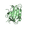
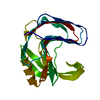
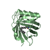
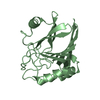
Title
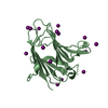
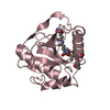
Crystal structure of peptidoglycan recognition protein-LE in complex with tracheal cytotoxin (monomeric diaminopimelic acid-type peptidoglycan)
ComponentsPEPTIDOGLYCAN-RECOGNITION PROTEIN-LE
KeywordsIMMUNE SYSTEM / PGRP / TRACHEAL CYTOTOXIN / INNATE IMMUNITY
Function / homology
Function and homology information


Peptidoglycans (PGN) bind to a peptidoglycan recognition protein receptor, PGRP-LC/LE / Peptidoglycan bound PGRP-LC/LE oligomerises / Assembly of the PGN:PGRP-LC/LE receptor 'signalling complex' / Activated IkappaB kinase (IKK) complex, Phospho IRD5:KEY dimer, phosphorylates REL in the PGN:PGRP-LC/LE receptor 'signalling complex' / REL binds to DREDD in the PGN:PGRP-LC/LE receptor 'signalling complex' / Phosphorylated REL is cleaved by and dissociates from DREDD / peptidoglycan recognition protein signaling pathway / Antimicrobial peptides / peptidoglycan immune receptor activity / Neutrophil degranulation ...Peptidoglycans (PGN) bind to a peptidoglycan recognition protein receptor, PGRP-LC/LE / Peptidoglycan bound PGRP-LC/LE oligomerises / Assembly of the PGN:PGRP-LC/LE receptor 'signalling complex' / Activated IkappaB kinase (IKK) complex, Phospho IRD5:KEY dimer, phosphorylates REL in the PGN:PGRP-LC/LE receptor 'signalling complex' / REL binds to DREDD in the PGN:PGRP-LC/LE receptor 'signalling complex' / Phosphorylated REL is cleaved by and dissociates from DREDD / peptidoglycan recognition protein signaling pathway / Antimicrobial peptides / peptidoglycan immune receptor activity / Neutrophil degranulation / peptidoglycan binding / positive regulation of innate immune response / positive regulation of phagocytosis / determination of adult lifespan / defense response to Gram-negative bacterium / defense response to virus / defense response to Gram-positive bacterium / defense response to bacterium / immune response / innate immune response / zinc ion binding / extracellular region / cytosol
Similarity search - Function
Peptidoglycan recognition protein family domain, metazoa/bacteria / Peptidoglycan recognition protein / Animal peptidoglycan recognition proteins homologous to Bacteriophage T3 lysozyme. / Lysozyme-like / Peptidoglycan recognition protein-like / Ami_2 / N-acetylmuramoyl-L-alanine amidase / N-acetylmuramoyl-L-alanine amidase domain / N-acetylmuramoyl-L-alanine amidase/PGRP domain superfamily / 3-Layer(aba) Sandwich / Alpha Beta
Similarity search - Domain/homology
Chem-MLD / Peptidoglycan-recognition protein LE
Similarity search - Component
Biological speciesDROSOPHILA MELANOGASTER (fruit fly)
MethodX-RAY DIFFRACTION / SYNCHROTRON / MOLECULAR REPLACEMENT / Resolution: 2.4 Å
AuthorsLim, J.-H. / Kim, M.-S. / Oh, B.-H.
CitationJournal: J.Biol.Chem. / Year: 2006
Title: Structural Basis for Preferential Recognition of Diaminopimelic Acid-Type Peptidoglycan by a Subset of Peptidoglycan Recognition Proteins
Authors: Lim, J.-H. / Kim, M.-S. / Kim, H.-E. / Yano, T. / Oshima, Y. / Aggarwal, K. / Goldman, W.E. / Silverman, N. / Kurata, S. / Oh, B.-H.
History
DepositionDec 29, 2005Deposition site: PDBE / Processing site: PDBE
Revision 1.0Jan 26, 2006Provider: repository / Type: Initial release
Revision 1.1May 8, 2011Group: Version format compliance
Revision 1.2Jul 13, 2011Group: Version format compliance
Revision 1.3Dec 13, 2023Group: Data collection / Database references ...Data collection / Database references / Other / Refinement description
Category: chem_comp_atom / chem_comp_bond ...chem_comp_atom / chem_comp_bond / database_2 / pdbx_database_status / pdbx_initial_refinement_model
Item: _database_2.pdbx_DOI / _database_2.pdbx_database_accession / _pdbx_database_status.status_code_sf
Remark 650 HELIX DETERMINATION METHOD: AUTHOR PROVIDED.
Remark 700 SHEET DETERMINATION METHOD: AUTHOR PROVIDED.

-
Structure visualization

Structure viewerMolecule:
MolmilJmol/JSmol

Downloads & links

-
Assembly

Deposited unit
A: PEPTIDOGLYCAN-RECOGNITION PROTEIN-LE
B: PEPTIDOGLYCAN-RECOGNITION PROTEIN-LE
C: PEPTIDOGLYCAN-RECOGNITION PROTEIN-LE
D: PEPTIDOGLYCAN-RECOGNITION PROTEIN-LE
hetero molecules


Theoretical massNumber of molelcules
Total (without water)83,9239
Polymers80,1434
Non-polymers3,7805
Water3,693205
1
A: PEPTIDOGLYCAN-RECOGNITION PROTEIN-LE
hetero molecules


Theoretical massNumber of molelcules
Total (without water)21,0503
Polymers20,0361
Non-polymers1,0142
Water181
TypeNameSymmetry operationNumber
identity operation1_555x,y,z1
MethodPQS
2
B: PEPTIDOGLYCAN-RECOGNITION PROTEIN-LE
hetero molecules


Theoretical massNumber of molelcules
Total (without water)20,9582
Polymers20,0361
Non-polymers9221
Water181
TypeNameSymmetry operationNumber
identity operation1_555x,y,z1
MethodPQS
3
C: PEPTIDOGLYCAN-RECOGNITION PROTEIN-LE
hetero molecules


Theoretical massNumber of molelcules
Total (without water)20,9582
Polymers20,0361
Non-polymers9221
Water181
TypeNameSymmetry operationNumber
identity operation1_555x,y,z1
MethodPQS
4
D: PEPTIDOGLYCAN-RECOGNITION PROTEIN-LE
hetero molecules


Theoretical massNumber of molelcules
Total (without water)20,9582
Polymers20,0361
Non-polymers9221
Water181
TypeNameSymmetry operationNumber
identity operation1_555x,y,z1
MethodPQS
Unit cell
Length a, b, c (Å)217.530, 217.530, 217.530
Angle α, β, γ (deg.)90.00, 90.00, 90.00
Int Tables number197
Space group name H-MI23
DetailsTHE FOUR MOLECULES IN THE ASYMMETRIC UNIT FORM A REPEATINGUNIT OF AN INFINITE FIBER.

-
Components

#1: Protein
PEPTIDOGLYCAN-RECOGNITION PROTEIN-LE


Mass: 20035.729 Da / Num. of mol.: 4 / Fragment: RESIDUES 173-345 / Mutation: YES
Source method: isolated from a genetically manipulated source
Details: PEPTIDOGLYCAN RECOGNITION PROTEIN-LE IN COMPLEX WITH TRACHEAL CYTOTOXIN
Source: (gene. exp.) DROSOPHILA MELANOGASTER (fruit fly) / Production host: ESCHERICHIA COLI (E. coli) / References: UniProt: Q9VXN9
#2: Chemical ChemComp-GOL / GLYCEROL / GLYCERIN / PROPANE-1,2,3-TRIOL


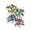
Mass: 92.094 Da / Num. of mol.: 1 / Source method: obtained synthetically / Formula: C3H8O3
#3: Chemical
ChemComp-MLD / GLCNAC(BETA1-4)-MURNAC(1,6-ANHYDRO)-L-ALA-GAMMA-D-GLU-MESO-A2PM-D-ALA / 2-ACETAMIDO-2-DEOXY-BETA-D-GLUCOPYRANOSE(BETA1-4)-2-ACETAMIDO-1,6-ANHYDRO-3-O-[(R)-1-CARBOXYETHYL]-2-DEOXY-BETA-D-GLUCOPYRANOSE-L-ALANYL-GAMMA-D-GLUTAMYL-MESO-DIAMINOPIMELYL-D-ALANINE


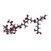
Mass: 921.899 Da / Num. of mol.: 4 / Source method: obtained synthetically / Formula: C37H59N7O20
#4: Water ChemComp-HOH / water


Mass: 18.015 Da / Num. of mol.: 205 / Source method: isolated from a natural source / Formula: H2O
Compound detailsENGINEERED RESIDUE IN CHAIN A, CYS 227 TO SER ENGINEERED RESIDUE IN CHAIN B, CYS 227 TO SER ...ENGINEERED RESIDUE IN CHAIN A, CYS 227 TO SER ENGINEERED RESIDUE IN CHAIN B, CYS 227 TO SER ENGINEERED RESIDUE IN CHAIN C, CYS 227 TO SER ENGINEERED RESIDUE IN CHAIN D, CYS 227 TO SER

-
Experimental details

-
Experiment

ExperimentMethod: X-RAY DIFFRACTION

-
Sample preparation

CrystalDensity Matthews: 5.16 Å3/Da / Density % sol: 75.96 %

-
Data collection

DiffractionMean temperature: 100 K
Diffraction sourceSource: SYNCHROTRON / Site: PAL/PLS / Beamline: 4A / Wavelength: 1
DetectorType: ADSC CCD / Detector: CCD
RadiationProtocol: SINGLE WAVELENGTH / Monochromatic (M) / Laue (L): M / Scattering type: x-ray
Radiation wavelengthWavelength: 1 Å / Relative weight: 1
ReflectionResolution: 2.4→30 Å / Num. obs: 66354 / % possible obs: 92.9 % / Observed criterion σ(I): 1 / Redundancy: 2.5 % / Rmerge(I) obs: 0.09 / Net I/σ(I): 24.3
Reflection shellResolution: 2.4→2.49 Å / Rmerge(I) obs: 0.2 / Mean I/σ(I) obs: 4.9 / % possible all: 90.4

-
Processing

SoftwareName: CNS / Version: 1.1 / Classification: refinement
RefinementMethod to determine structure: MOLECULAR REPLACEMENT
Starting model: PDB ENTRY 1OHT
Resolution: 2.4→30 Å / Cross valid method: THROUGHOUT / σ(F): 1
RfactorNum. reflection% reflectionSelection details
Rfree0.2145 3103 5 %RANDOM
Rwork0.1985 ---
obs0.1985 61492 92.3 %-
Solvent computationBsol: 28.5351 Å2 / ksol: 0.374375 e/Å3
Refinement stepCycle: LAST / Resolution: 2.4→30 Å
ProteinNucleic acidLigandSolventTotal
Num. atoms5475 0 262 205 5942
Refine LS restraints
Refine-IDTypeDev ideal
X-RAY DIFFRACTIONc_bond_d0.005986
X-RAY DIFFRACTIONc_bond_d_na
X-RAY DIFFRACTIONc_bond_d_prot
X-RAY DIFFRACTIONc_angle_d
X-RAY DIFFRACTIONc_angle_d_na
X-RAY DIFFRACTIONc_angle_d_prot
X-RAY DIFFRACTIONc_angle_deg1.30458
X-RAY DIFFRACTIONc_angle_deg_na
X-RAY DIFFRACTIONc_angle_deg_prot
X-RAY DIFFRACTIONc_dihedral_angle_d
X-RAY DIFFRACTIONc_dihedral_angle_d_na
X-RAY DIFFRACTIONc_dihedral_angle_d_prot
X-RAY DIFFRACTIONc_improper_angle_d
X-RAY DIFFRACTIONc_improper_angle_d_na
X-RAY DIFFRACTIONc_improper_angle_d_prot
X-RAY DIFFRACTIONc_mcbond_it
X-RAY DIFFRACTIONc_mcangle_it
X-RAY DIFFRACTIONc_scbond_it
X-RAY DIFFRACTIONc_scangle_it
Xplor file
Refine-IDSerial noParam file
X-RAY DIFFRACTION1PROTEIN_REP.PARAM
X-RAY DIFFRACTION2WATER.PARAM
X-RAY DIFFRACTION3ION.PARAM
X-RAY DIFFRACTION4MLD.PAR
X-RAY DIFFRACTION5GLY.PAR

+
About Yorodumi

-
News

-
Feb 9, 2022. New format data for meta-information of EMDB entries

New format data for meta-information of EMDB entries

  • Version 3 of the EMDB header file is now the official format.
  • The previous official version 1.9 will be removed from the archive.

Related info.:EMDB header

External links:wwPDB to switch to version 3 of the EMDB data model

-
Aug 12, 2020. Covid-19 info

Covid-19 info

URL: https://pdbjlvh1.pdbj.org/emnavi/covid19.php

New page: Covid-19 featured information page in EM Navigator.

Related info.:Covid-19 info / Mar 5, 2020. Novel coronavirus structure data

+
Mar 5, 2020. Novel coronavirus structure data

Novel coronavirus structure data

Related info.:Yorodumi Speices / Aug 12, 2020. Covid-19 info

External links:COVID-19 featured content - PDBj / Molecule of the Month (242):Coronavirus Proteases

+
Jan 31, 2019. EMDB accession codes are about to change! (news from PDBe EMDB page)

EMDB accession codes are about to change! (news from PDBe EMDB page)

  • The allocation of 4 digits for EMDB accession codes will soon come to an end. Whilst these codes will remain in use, new EMDB accession codes will include an additional digit and will expand incrementally as the available range of codes is exhausted. The current 4-digit format prefixed with “EMD-” (i.e. EMD-XXXX) will advance to a 5-digit format (i.e. EMD-XXXXX), and so on. It is currently estimated that the 4-digit codes will be depleted around Spring 2019, at which point the 5-digit format will come into force.
  • The EM Navigator/Yorodumi systems omit the EMD- prefix.

Related info.:Q: What is EMD? / ID/Accession-code notation in Yorodumi/EM Navigator

External links:EMDB Accession Codes are Changing Soon! / Contact to PDBj

+
Jul 12, 2017. Major update of PDB

Major update of PDB

  • wwPDB released updated PDB data conforming to the new PDBx/mmCIF dictionary.
  • This is a major update changing the version number from 4 to 5, and with Remediation, in which all the entries are updated.
  • In this update, many items about electron microscopy experimental information are reorganized (e.g. em_software).
  • Now, EM Navigator and Yorodumi are based on the updated data.

External links:wwPDB Remediation / Enriched Model Files Conforming to OneDep Data Standards Now Available in the PDB FTP Archive

-
Yorodumi

Thousand views of thousand structures

  • Yorodumi is a browser for structure data from EMDB, PDB, SASBDB, etc.
  • This page is also the successor to EM Navigator detail page, and also detail information page/front-end page for Omokage search.
  • The word "yorodu" (or yorozu) is an old Japanese word meaning "ten thousand". "mi" (miru) is to see.

Related info.:EMDB / PDB / SASBDB / Comparison of 3 databanks / Yorodumi Search / Aug 31, 2016. New EM Navigator & Yorodumi / Yorodumi Papers / Jmol/JSmol / Function and homology information / Changes in new EM Navigator and Yorodumi

Read more