 Movie
Movie Controller

[English] 日本語
 Yorodumi
- PDB-1hv1: DISSECTING ELECTROSTATIC INTERACTIONS AND THE PH-DEPENDENT ACTIVI... -
+ Open data

- Basic information
| Entry | Database: PDB / ID: 1hv1 | ||||||
|---|---|---|---|---|---|---|---|
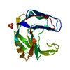
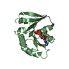
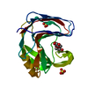
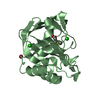
| Title | DISSECTING ELECTROSTATIC INTERACTIONS AND THE PH-DEPENDENT ACTIVITY OF A FAMILY 11 GLYCOSIDASE | ||||||
 Components Components | ENDO-1,4-BETA-XYLANASE | ||||||
 Keywords Keywords | HYDROLASE / beta sheet | ||||||
| Function / homology |  Function and homology information Function and homology informationendo-1,4-beta-xylanase / endo-1,4-beta-xylanase activity / xylan catabolic process Similarity search - Function | ||||||
| Biological species |  Bacillus circulans (bacteria) Bacillus circulans (bacteria) | ||||||
| Method |  X-RAY DIFFRACTION / MOLECULAR REPLACEMENT / Resolution: 1.8 Å X-RAY DIFFRACTION / MOLECULAR REPLACEMENT / Resolution: 1.8 Å | ||||||
 Authors Authors | Joshi, M.D. / Sidhu, G. / Nielsen, J.E. / Brayer, G.D. / Withers, S.G. / McIntosh, L.P. | ||||||
 Citation Citation | Journal: Biochemistry / Year: 2001 Title: Dissecting the electrostatic interactions and pH-dependent activity of a family 11 glycosidase. Authors: Joshi, M.D. / Sidhu, G. / Nielsen, J.E. / Brayer, G.D. / Withers, S.G. / McIntosh, L.P. | ||||||
| History |
|
- Structure visualization
| Structure viewer | Molecule: MolmilJmol/JSmol |
|---|
- Downloads & links
-Download
| PDBx/mmCIF format | 1hv1.cif.gz | 51.4 KB | Display | PDBx/mmCIF format |
|---|---|---|---|---|
| PDB format | pdb1hv1.ent.gz | 36.2 KB | Display | PDB format |
| PDBx/mmJSON format | 1hv1.json.gz | Tree view | PDBx/mmJSON format | |
| Others |  Other downloads Other downloads |
-Validation report
| Arichive directory | https://data.pdbj.org/pub/pdb/validation_reports/hv/1hv1ftp://data.pdbj.org/pub/pdb/validation_reports/hv/1hv1 | HTTPS FTP |
|---|
-Related structure data
| Related structure data |  1hv0C  1bcxS S: Starting model for refinement C: citing same article ( |
|---|---|
| Similar structure data |


-Links
 PDBj
PDBj
- Assembly
| Deposited unit | 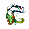
| ||||||||||
|---|---|---|---|---|---|---|---|---|---|---|---|
| 1 |
| ||||||||||
| Unit cell |
|
-Components
| #1: Protein | Mass: 20351.955 Da / Num. of mol.: 1 / Fragment: Q127A_BCX / Mutation: Q127A Source method: isolated from a genetically manipulated source Source: (gene. exp.) Bacillus circulans (bacteria) / Plasmid: PCW / Species (production host): Escherichia coli / Production host: |
|---|---|
| #2: Water | ChemComp-HOH /  Mass: 18.015 Da / Num. of mol.: 177 / Source method: isolated from a natural source / Formula: H2O Mass: 18.015 Da / Num. of mol.: 177 / Source method: isolated from a natural source / Formula: H2O |
-Experimental details
-Experiment
| Experiment | Method: X-RAY DIFFRACTION / Number of used crystals: 1 |
|---|
- Sample preparation
| Crystal | Density Matthews: 2.23 Å3/Da / Density % sol: 44.77 % | ||||||||||||||||||||||||||||||
|---|---|---|---|---|---|---|---|---|---|---|---|---|---|---|---|---|---|---|---|---|---|---|---|---|---|---|---|---|---|---|---|
| Crystal grow | Temperature: 298 K / Method: vapor diffusion, hanging drop / pH: 7.5 Details: ammonimu sulphate, sodium chloride, TRIS, pH 7.5, VAPOR DIFFUSION, HANGING DROP at 298 K | ||||||||||||||||||||||||||||||
| Crystal grow | *PLUS Details: Sidhu, G., (1999) Biochemistry, 38, 5346. | ||||||||||||||||||||||||||||||
| Components of the solutions | *PLUS
|
-Data collection
| Diffraction | Mean temperature: 298 K |
|---|---|
| Diffraction source | Source: ROTATING ANODE / Type: RIGAKU RU300 / Wavelength: 1.5418 Å |
| Detector | Type: RIGAKU RAXIS IIC / Detector: IMAGE PLATE / Date: Oct 1, 1999 |
| Radiation | Monochromator: graphite / Protocol: SINGLE WAVELENGTH / Monochromatic (M) / Laue (L): M / Scattering type: x-ray |
| Radiation wavelength | Wavelength: 1.5418 Å / Relative weight: 1 |
| Reflection | Resolution: 1.8→50 Å / Num. obs: 17548 / Observed criterion σ(F): 0 / Observed criterion σ(I): 0 / Redundancy: 9 % / Rmerge(I) obs: 0.054 / Net I/σ(I): 21.8 |
| Reflection shell | Resolution: 1.8→1.88 Å / Rmerge(I) obs: 0.115 |
| Reflection | *PLUS Lowest resolution: 9999 Å / Num. measured all: 157188 |
| Reflection shell | *PLUS Mean I/σ(I) obs: 6.6 |
- Processing
| Software |
| ||||||||||||||||||
|---|---|---|---|---|---|---|---|---|---|---|---|---|---|---|---|---|---|---|---|
| Refinement | Method to determine structure: MOLECULAR REPLACEMENT Starting model: PDB ENTRY 1BCX Resolution: 1.8→10 Å / σ(F): 0
| ||||||||||||||||||
| Refinement step | Cycle: LAST / Resolution: 1.8→10 Å
| ||||||||||||||||||
| Refine LS restraints |
| ||||||||||||||||||
| LS refinement shell | Resolution: 1.8→10 Å
| ||||||||||||||||||
| Software | *PLUS Name: X-PLOR / Version: 3.851 / Classification: refinement | ||||||||||||||||||
| Refinement | *PLUS Highest resolution: 1.8 Å / Lowest resolution: 10 Å / σ(F): 0 | ||||||||||||||||||
| Solvent computation | *PLUS | ||||||||||||||||||
| Displacement parameters | *PLUS | ||||||||||||||||||
| LS refinement shell | *PLUS Highest resolution: 1.8 Å / Lowest resolution: 10 Å / Rfactor Rwork: 0.164 |
