 Movie
Movie Controller


+ Open data

- Basic information
| Entry | Database: PDB / ID: 6vri | ||||||
|---|---|---|---|---|---|---|---|
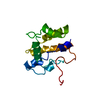
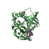
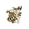
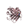
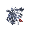
| Title | Crystal Structure of the wtBlc-split Protein | ||||||
 Components Components | (Outer membrane lipoprotein Blc) x 2 | ||||||
 Keywords Keywords | UNKNOWN FUNCTION / lipocalin / heme / beta barrel / split protein | ||||||
| Function / homology |  Function and homology information Function and homology informationresponse to stress / cell outer membrane / DNA damage response / lipid binding Similarity search - Function | ||||||
| Biological species |  | ||||||
| Method |  X-RAY DIFFRACTION / SYNCHROTRON / MOLECULAR REPLACEMENT / molecular replacement / Resolution: 1.94 Å X-RAY DIFFRACTION / SYNCHROTRON / MOLECULAR REPLACEMENT / molecular replacement / Resolution: 1.94 Å | ||||||
 Authors Authors | Bozhanova, N.G. / Meiler, J. | ||||||
| Funding support |  United States, 1items United States, 1items
| ||||||
 Citation Citation | Journal: Febs Lett. / Year: 2021 Title: Lipocalin Blc is a potential heme-binding protein. Authors: Bozhanova, N.G. / Calcutt, M.W. / Beavers, W.N. / Brown, B.P. / Skaar, E.P. / Meiler, J. | ||||||
| History |
|
- Structure visualization
| Structure viewer | Molecule: MolmilJmol/JSmol |
|---|
- Downloads & links
-Download
| PDBx/mmCIF format | 6vri.cif.gz | 111.5 KB | Display | PDBx/mmCIF format |
|---|---|---|---|---|
| PDB format | pdb6vri.ent.gz | 83.9 KB | Display | PDB format |
| PDBx/mmJSON format | 6vri.json.gz | Tree view | PDBx/mmJSON format | |
| Others |  Other downloads Other downloads |
-Validation report
| Arichive directory | https://data.pdbj.org/pub/pdb/validation_reports/vr/6vriftp://data.pdbj.org/pub/pdb/validation_reports/vr/6vri | HTTPS FTP |
|---|
-Related structure data
| Related structure data | 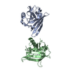 1qwdS S: Starting model for refinement |
|---|---|
| Similar structure data |


-Links
 PDBj
PDBj


- Assembly
| Deposited unit | 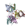
| |||||||||
|---|---|---|---|---|---|---|---|---|---|---|
| 1 | 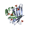
| |||||||||
| 2 | 
| |||||||||
| 3 | 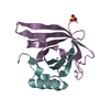
| |||||||||
| Unit cell |
| |||||||||
| Components on special symmetry positions |
|
-Components
| #1: Protein | Mass: 12266.708 Da / Num. of mol.: 3 / Fragment: N-terminal fragment (UNP residues 23-109) Source method: isolated from a genetically manipulated source Source: (gene. exp.) #2: Protein | Mass: 7902.951 Da / Num. of mol.: 3 / Fragment: C-terminal fragment (UNP residues 110-177) Source method: isolated from a genetically manipulated source Source: (gene. exp.) #3: Chemical | ChemComp-SO4 /   Mass: 96.063 Da / Num. of mol.: 13 / Source method: obtained synthetically / Formula: SO4 Mass: 96.063 Da / Num. of mol.: 13 / Source method: obtained synthetically / Formula: SO4#4: Chemical | ChemComp-HEM / |   Mass: 616.487 Da / Num. of mol.: 1 / Source method: obtained synthetically / Formula: C34H32FeN4O4 / Feature type: SUBJECT OF INVESTIGATION Mass: 616.487 Da / Num. of mol.: 1 / Source method: obtained synthetically / Formula: C34H32FeN4O4 / Feature type: SUBJECT OF INVESTIGATION#5: Water | ChemComp-HOH / |  Mass: 18.015 Da / Num. of mol.: 72 / Source method: isolated from a natural source / Formula: H2O Mass: 18.015 Da / Num. of mol.: 72 / Source method: isolated from a natural source / Formula: H2OHas ligand of interest | Y | |
|---|
-Experimental details
-Experiment
| Experiment | Method: X-RAY DIFFRACTION / Number of used crystals: 1 |
|---|
- Sample preparation
| Crystal | Density Matthews: 2.43 Å3/Da / Density % sol: 49.41 % / Mosaicity: 0.217 ° |
|---|---|
| Crystal grow | Temperature: 294 K / Method: vapor diffusion, hanging drop / pH: 4.5 Details: 1.6 M ammonium sulfate, 0.1 M MES, pH 4.5, supplemented with 5% w/v n-Dodecyl-b-D-maltoside |
-Data collection
| Diffraction | Mean temperature: 100 K / Serial crystal experiment: N |
|---|---|
| Diffraction source | Source: SYNCHROTRON / Site: APS / Beamline: 21-ID-F / Wavelength: 0.97872 Å |
| Detector | Type: MARMOSAIC 225 mm CCD / Detector: CCD / Date: Dec 16, 2018 |
| Radiation | Monochromator: diamond(111) / Protocol: SINGLE WAVELENGTH / Monochromatic (M) / Laue (L): M / Scattering type: x-ray |
| Radiation wavelength | Wavelength: 0.97872 Å / Relative weight: 1 |
| Reflection | Resolution: 1.94→59.25 Å / Num. obs: 44856 / % possible obs: 99.5 % / Redundancy: 9 % / Biso Wilson estimate: 30.626 Å2 / Rpim(I) all: 0.051 / Rrim(I) all: 0.15 / Net I/σ(I): 8.1 / Num. measured all: 403082 |
| Reflection shell | Resolution: 1.94→1.97 Å / Redundancy: 8.9 % / Mean I/σ(I) obs: 2.1 / Num. unique obs: 1915 / Rpim(I) all: 0.534 / Rrim(I) all: 1.627 / % possible all: 86.2 |
-Phasing
| Phasing | Method: molecular replacement | ||||||
|---|---|---|---|---|---|---|---|
| Phasing MR | R rigid body: 0.414
|
- Processing
| Software |
| |||||||||||||||||||||||||||||||||||||||||||||||||||||||||||||||||||||||||||
|---|---|---|---|---|---|---|---|---|---|---|---|---|---|---|---|---|---|---|---|---|---|---|---|---|---|---|---|---|---|---|---|---|---|---|---|---|---|---|---|---|---|---|---|---|---|---|---|---|---|---|---|---|---|---|---|---|---|---|---|---|---|---|---|---|---|---|---|---|---|---|---|---|---|---|---|---|
| Refinement | Method to determine structure: MOLECULAR REPLACEMENT Starting model: PDB entry 1QWD Resolution: 1.94→59.25 Å / Cor.coef. Fo:Fc: 0.947 / Cor.coef. Fo:Fc free: 0.934 / Cross valid method: THROUGHOUT / σ(F): 0 / ESU R: 0.159 / ESU R Free: 0.149 / Stereochemistry target values: MAXIMUM LIKELIHOOD Details: HYDROGENS HAVE BEEN ADDED IN THE RIDING POSITIONS U VALUES : REFINED INDIVIDUALLY
| |||||||||||||||||||||||||||||||||||||||||||||||||||||||||||||||||||||||||||
| Solvent computation | Ion probe radii: 0.8 Å / Shrinkage radii: 0.8 Å / VDW probe radii: 1.2 Å / Solvent model: MASK | |||||||||||||||||||||||||||||||||||||||||||||||||||||||||||||||||||||||||||
| Displacement parameters | Biso max: 104.16 Å2 / Biso mean: 32.65 Å2 / Biso min: 16.8 Å2
| |||||||||||||||||||||||||||||||||||||||||||||||||||||||||||||||||||||||||||
| Refinement step | Cycle: final / Resolution: 1.94→59.25 Å
| |||||||||||||||||||||||||||||||||||||||||||||||||||||||||||||||||||||||||||
| Refine LS restraints |
| |||||||||||||||||||||||||||||||||||||||||||||||||||||||||||||||||||||||||||
| LS refinement shell | Resolution: 1.94→1.987 Å / Rfactor Rfree error: 0
|
