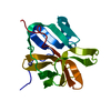
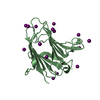
protein transport by the Sec complex / protein secretion by the type II secretion system / Hydrolases; Acting on peptide bonds (peptidases); Metalloendopeptidases / peptidoglycan catabolic process / metalloendopeptidase activity / endopeptidase activity / proteolysis / : / metal ion binding Similarity search - Function
Redundancy: 3.5 % / Av σ(I) over netI: 23.39 / Number: 109259 / Rmerge(I) obs: 0.095 / Χ2: 1.06 / D res high: 2.14 Å / D res low: 50 Å / Num. obs: 31654 / % possible obs: 99.7
Diffraction reflection shell
Highest resolution (Å)
Lowest resolution (Å)
% possible obs (%)
ID
Rmerge(I) obs
Chi squared
Redundancy
4.61
50
100
1
0.045
1.073
3.8
3.66
4.61
100
1
0.047
1.063
3.7
3.2
3.66
100
1
0.06
0.999
3.7
2.9
3.2
100
1
0.088
0.977
3.7
2.7
2.9
100
1
0.13
0.984
3.6
2.54
2.7
100
1
0.186
1.017
3.6
2.41
2.54
100
1
0.229
1.066
3.5
2.31
2.41
100
1
0.261
1.129
3.3
2.22
2.31
99.5
1
0.278
1.179
3
2.14
2.22
98
1
0.299
1.21
2.6
Reflection
Resolution: 2.14→50 Å / Num. obs: 32280 / % possible obs: 100 % / Redundancy: 3.7 % / Rmerge(I) obs: 0.054 / Χ2: 1.027 / Net I/σ(I): 14.7
Reflection shell
Resolution (Å)
Redundancy (%)
Rmerge(I) obs
Num. unique all
Χ2
Diffraction-ID
% possible all
2.14-2.22
3.6
0.1
3264
1.063
1
100
2.22-2.31
3.7
0.095
3216
1.088
1
100
2.31-2.41
3.7
0.086
3203
1.04
1
100
2.41-2.54
3.7
0.079
3261
1.065
1
100
2.54-2.7
3.7
0.071
3217
1.031
1
100
2.7-2.9
3.7
0.06
3209
0.992
1
100
2.9-3.2
3.7
0.05
3222
0.994
1
100
3.2-3.66
3.7
0.042
3216
1.038
1
100
3.66-4.61
3.7
0.037
3231
0.965
1
100
4.61-50
3.7
0.037
3241
1
1
99.9
-
Phasing
Phasing
Method: MAD
-
Processing
Software
Name
Version
Classification
NB
DENZO
datareduction
SCALEPACK
datascaling
SOLVE
phasing
RESOLVE
phasing
REFMAC
refinement
PDB_EXTRACT
3.005
dataextraction
Refinement
Method to determine structure: MAD / Resolution: 2.14→37.57 Å / Cor.coef. Fo:Fc: 0.952 / Cor.coef. Fo:Fc free: 0.919 / WRfactor Rfree: 0.189 / WRfactor Rwork: 0.145 / Occupancy max: 1 / Occupancy min: 0.5 / FOM work R set: 0.891 / SU B: 3.684 / SU ML: 0.098 / SU R Cruickshank DPI: 0.322 / SU Rfree: 0.189 / Cross valid method: THROUGHOUT / σ(F): 0 / ESU R: 0.322 / ESU R Free: 0.189 / Stereochemistry target values: MAXIMUM LIKELIHOOD Details: HYDROGENS HAVE BEEN ADDED IN THE RIDING POSITIONS. U VALUES: REFINED INDIVIDUALLY
Rfactor
Num. reflection
% reflection
Selection details
Rfree
0.195
882
5.1 %
RANDOM
Rwork
0.149
-
-
-
obs
0.151
17436
99.98 %
-
Solvent computation
Ion probe radii: 0.8 Å / Shrinkage radii: 0.8 Å / VDW probe radii: 1.4 Å / Solvent model: MASK
In the structure databanks used in Yorodumi, some data are registered as the other names, "COVID-19 virus" and "2019-nCoV". Here are the details of the virus and the list of structure data.
Jan 31, 2019. EMDB accession codes are about to change! (news from PDBe EMDB page)
EMDB accession codes are about to change! (news from PDBe EMDB page)
The allocation of 4 digits for EMDB accession codes will soon come to an end. Whilst these codes will remain in use, new EMDB accession codes will include an additional digit and will expand incrementally as the available range of codes is exhausted. The current 4-digit format prefixed with “EMD-” (i.e. EMD-XXXX) will advance to a 5-digit format (i.e. EMD-XXXXX), and so on. It is currently estimated that the 4-digit codes will be depleted around Spring 2019, at which point the 5-digit format will come into force.
The EM Navigator/Yorodumi systems omit the EMD- prefix.
Related info.:Q: What is EMD? / ID/Accession-code notation in Yorodumi/EM Navigator
Yorodumi is a browser for structure data from EMDB, PDB, SASBDB, etc.
This page is also the successor to EM Navigator detail page, and also detail information page/front-end page for Omokage search.
The word "yorodu" (or yorozu) is an old Japanese word meaning "ten thousand". "mi" (miru) is to see.
Related info.:EMDB / PDB / SASBDB / Comparison of 3 databanks / Yorodumi Search / Aug 31, 2016. New EM Navigator & Yorodumi / Yorodumi Papers / Jmol/JSmol / Function and homology information / Changes in new EM Navigator and Yorodumi
 Movie
Movie Controller

 Yorodumi
 Open data
 Basic information
 Components
 Keywords
 Function and homology information
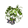
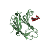
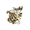
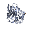
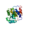
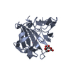
 Pseudomonas aeruginosa (bacteria)
 X-RAY DIFFRACTION /
  Authors
 Citation
 Structure visualization
 Downloads & links
 Other downloads


 PDBj
PDBj
 Assembly
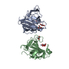


 Mass: 65.409 Da / Num. of mol.: 2 / Source method: obtained synthetically / Formula: Zn
 Mass: 150.087 Da / Num. of mol.: 2 / Source method: obtained synthetically / Formula: C4H6O6
 Mass: 92.094 Da / Num. of mol.: 3 / Source method: obtained synthetically / Formula: C3H8O3
 Mass: 18.015 Da / Num. of mol.: 391 / Source method: isolated from a natural source / Formula: H2O
 Sample preparation
 / Beamline: 19-ID / Wavelength: 1.28402, 1.28348, 1.2574
 Processing