 Movie
Movie Controller

[English] 日本語
 Yorodumi
- PDB-5hzj: Crystal structure of photoinhibitable Intersectin1 containing wil... -
+ Open data

- Basic information
| Entry | Database: PDB / ID: 5hzj | ||||||
|---|---|---|---|---|---|---|---|
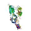
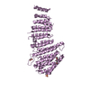
| Title | Crystal structure of photoinhibitable Intersectin1 containing wildtype LOV2 domain | ||||||
 Components Components | Intersectin-1,NPH1-1,Intersectin-1 | ||||||
 Keywords Keywords | SIGNALING PROTEIN / Photoswitch / Chimera | ||||||
| Function / homology |  Function and homology information Function and homology informationclathrin-dependent synaptic vesicle endocytosis / positive regulation of caveolin-mediated endocytosis / positive regulation of growth hormone secretion / postsynaptic endocytic zone / blue light photoreceptor activity / regulation of modification of postsynaptic actin cytoskeleton / proline-rich region binding / postsynaptic actin cytoskeleton / apical dendrite / regulation of small GTPase mediated signal transduction ...clathrin-dependent synaptic vesicle endocytosis / positive regulation of caveolin-mediated endocytosis / positive regulation of growth hormone secretion / postsynaptic endocytic zone / blue light photoreceptor activity / regulation of modification of postsynaptic actin cytoskeleton / proline-rich region binding / postsynaptic actin cytoskeleton / apical dendrite / regulation of small GTPase mediated signal transduction / regulation of postsynapse organization / endosomal transport / NRAGE signals death through JNK / RHOQ GTPase cycle / positive regulation of dendritic spine development / intracellular vesicle / exocytosis / CDC42 GTPase cycle / RHOG GTPase cycle / EPHB-mediated forward signaling / clathrin-coated pit / guanyl-nucleotide exchange factor activity / recycling endosome / intracellular protein localization / nuclear envelope / Cargo recognition for clathrin-mediated endocytosis / protein transport / lamellipodium / G alpha (12/13) signalling events / Clathrin-mediated endocytosis / presynaptic membrane / molecular adaptor activity / dendritic spine / protein-macromolecule adaptor activity / non-specific serine/threonine protein kinase / intracellular signal transduction / protein serine/threonine kinase activity / neuronal cell body / calcium ion binding / glutamatergic synapse / ATP binding / metal ion binding / plasma membrane / cytoplasm / cytosol Similarity search - Function | ||||||
| Biological species |  Homo sapiens (human) Homo sapiens (human) | ||||||
| Method |  X-RAY DIFFRACTION / SYNCHROTRON / MOLECULAR REPLACEMENT / Resolution: 2.6 Å X-RAY DIFFRACTION / SYNCHROTRON / MOLECULAR REPLACEMENT / Resolution: 2.6 Å | ||||||
 Authors Authors | Tarnawski, M. / Dagliyan, O. / Chu, P.H. / Shirvanyants, D. / Dokholyan, N.V. / Hahn, K.M. / Schlichting, I. | ||||||
 Citation Citation | Journal: Science / Year: 2016 Title: Engineering extrinsic disorder to control protein activity in living cells. Authors: Dagliyan, O. / Tarnawski, M. / Chu, P.H. / Shirvanyants, D. / Schlichting, I. / Dokholyan, N.V. / Hahn, K.M. | ||||||
| History |
|
- Structure visualization
| Structure viewer | Molecule: MolmilJmol/JSmol |
|---|
- Downloads & links
-Download
| PDBx/mmCIF format | 5hzj.cif.gz | 405.8 KB | Display | PDBx/mmCIF format |
|---|---|---|---|---|
| PDB format | pdb5hzj.ent.gz | 333.1 KB | Display | PDB format |
| PDBx/mmJSON format | 5hzj.json.gz | Tree view | PDBx/mmJSON format | |
| Others |  Other downloads Other downloads |
-Validation report
| Arichive directory | https://data.pdbj.org/pub/pdb/validation_reports/hz/5hzjftp://data.pdbj.org/pub/pdb/validation_reports/hz/5hzj | HTTPS FTP |
|---|
-Related structure data
| Related structure data | 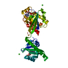 5hzhC 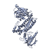 5hziC 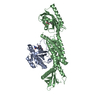 5hzkC 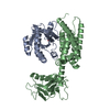 1ki1S 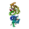 2wkqS S: Starting model for refinement C: citing same article ( |
|---|---|
| Similar structure data |


-Links
 PDBj
PDBj


- Assembly
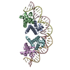
| Deposited unit | 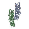
| ||||||||
|---|---|---|---|---|---|---|---|---|---|
| 1 | 
| ||||||||
| 2 | 
| ||||||||
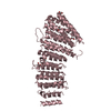
| Unit cell |
|
-Components
| #1: Protein | Mass: 58281.121 Da / Num. of mol.: 2 Source method: isolated from a genetically manipulated source Source: (gene. exp.) Homo sapiens (human), (gene. exp.) Gene: ITSN1, ITSN, SH3D1A, NPH1-1 / Production host:  #2: Chemical |   Mass: 456.344 Da / Num. of mol.: 2 / Source method: obtained synthetically / Formula: C17H21N4O9P Mass: 456.344 Da / Num. of mol.: 2 / Source method: obtained synthetically / Formula: C17H21N4O9P#3: Water | ChemComp-HOH / |  Mass: 18.015 Da / Num. of mol.: 75 / Source method: isolated from a natural source / Formula: H2O Mass: 18.015 Da / Num. of mol.: 75 / Source method: isolated from a natural source / Formula: H2O |
|---|
-Experimental details
-Experiment
| Experiment | Method: X-RAY DIFFRACTION / Number of used crystals: 1 |
|---|
- Sample preparation
| Crystal | Density Matthews: 2.59 Å3/Da / Density % sol: 52.56 % |
|---|---|
| Crystal grow | Temperature: 293 K / Method: vapor diffusion Details: 0.1 M HEPES pH 7.5, 9% (w/v) PEG 8000, 9% (v/v) ethylene glycol |
-Data collection
| Diffraction | Mean temperature: 100 K |
|---|---|
| Diffraction source | Source: SYNCHROTRON / Site: SLS  / Beamline: X10SA / Wavelength: 0.97903 Å / Beamline: X10SA / Wavelength: 0.97903 Å |
| Detector | Type: DECTRIS PILATUS 6M / Detector: PIXEL / Date: Oct 27, 2014 |
| Radiation | Monochromator: Si(111) / Protocol: SINGLE WAVELENGTH / Monochromatic (M) / Laue (L): M / Scattering type: x-ray |
| Radiation wavelength | Wavelength: 0.97903 Å / Relative weight: 1 |
| Reflection | Resolution: 2.6→50 Å / Num. obs: 35551 / % possible obs: 97 % / Redundancy: 3.4 % / Rmerge(I) obs: 0.059 / Net I/σ(I): 12.5 |
| Reflection shell | Resolution: 2.6→2.7 Å / Redundancy: 3.5 % / Rmerge(I) obs: 0.453 / Mean I/σ(I) obs: 2.6 / % possible all: 97.2 |
- Processing
| Software |
| |||||||||||||||||||||||||||||||||||||||||||||||||||||||||||||||||||||||||||||||||||||||||||||||||||||||||||||||||||||||||||||
|---|---|---|---|---|---|---|---|---|---|---|---|---|---|---|---|---|---|---|---|---|---|---|---|---|---|---|---|---|---|---|---|---|---|---|---|---|---|---|---|---|---|---|---|---|---|---|---|---|---|---|---|---|---|---|---|---|---|---|---|---|---|---|---|---|---|---|---|---|---|---|---|---|---|---|---|---|---|---|---|---|---|---|---|---|---|---|---|---|---|---|---|---|---|---|---|---|---|---|---|---|---|---|---|---|---|---|---|---|---|---|---|---|---|---|---|---|---|---|---|---|---|---|---|---|---|---|
| Refinement | Method to determine structure: MOLECULAR REPLACEMENT Starting model: 1KI1, 2WKQ Resolution: 2.6→47.024 Å / SU ML: 0.46 / Cross valid method: FREE R-VALUE / σ(F): 1.36 / Phase error: 34.74
| |||||||||||||||||||||||||||||||||||||||||||||||||||||||||||||||||||||||||||||||||||||||||||||||||||||||||||||||||||||||||||||
| Solvent computation | Shrinkage radii: 0.9 Å / VDW probe radii: 1.11 Å | |||||||||||||||||||||||||||||||||||||||||||||||||||||||||||||||||||||||||||||||||||||||||||||||||||||||||||||||||||||||||||||
| Refinement step | Cycle: LAST / Resolution: 2.6→47.024 Å
| |||||||||||||||||||||||||||||||||||||||||||||||||||||||||||||||||||||||||||||||||||||||||||||||||||||||||||||||||||||||||||||
| Refine LS restraints |
| |||||||||||||||||||||||||||||||||||||||||||||||||||||||||||||||||||||||||||||||||||||||||||||||||||||||||||||||||||||||||||||
| LS refinement shell |
| |||||||||||||||||||||||||||||||||||||||||||||||||||||||||||||||||||||||||||||||||||||||||||||||||||||||||||||||||||||||||||||
| Refinement TLS params. | Method: refined / Refine-ID: X-RAY DIFFRACTION
| |||||||||||||||||||||||||||||||||||||||||||||||||||||||||||||||||||||||||||||||||||||||||||||||||||||||||||||||||||||||||||||
| Refinement TLS group |
|
