 ムービー
ムービー コントローラー


+ データを開く

- 基本情報
基本情報
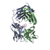
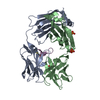
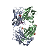
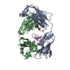
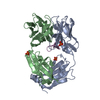
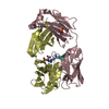
| 登録情報 | データベース: PDB / ID: 5hyq | ||||||
|---|---|---|---|---|---|---|---|
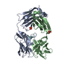
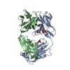
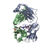
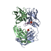
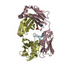
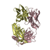
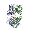
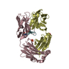
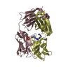
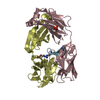
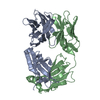
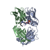
| タイトル | Cetuximab Fab in complex with amidated meditope | ||||||
 要素 要素 |
| ||||||
 キーワード キーワード | IMMUNE SYSTEM / antibody / anti-EGFR | ||||||
| 機能・相同性 | Immunoglobulins / Immunoglobulin-like / Sandwich / Mainly Beta / PHOSPHATE ION 機能・相同性情報 機能・相同性情報 | ||||||
| 生物種 |   HOMO SAPIENS (ヒト) HOMO SAPIENS (ヒト)synthetic construct (人工物) | ||||||
| 手法 |  X線回折 / 分子置換 / 解像度: 2.477 Å X線回折 / 分子置換 / 解像度: 2.477 Å | ||||||
 データ登録者 データ登録者 | Bzymek, K.P. / Williams, J.C. | ||||||
 引用 引用 | ジャーナル: Acta Crystallogr F Struct Biol Commun / 年: 2016 タイトル: Cyclization strategies of meditopes: affinity and diffraction studies of meditope-Fab complexes. 著者: Bzymek, K.P. / Ma, Y. / Avery, K.A. / Horne, D.A. / Williams, J.C. | ||||||
| 履歴 |
|
- 構造の表示
構造の表示
| 構造ビューア | 分子: MolmilJmol/JSmol |
|---|
- ダウンロードとリンク
-ダウンロード
| PDBx/mmCIF形式 | 5hyq.cif.gz | 190.8 KB | 表示 | PDBx/mmCIF形式 |
|---|---|---|---|---|
| PDB形式 | pdb5hyq.ent.gz | 149.9 KB | 表示 | PDB形式 |
| PDBx/mmJSON形式 | 5hyq.json.gz | ツリー表示 | PDBx/mmJSON形式 | |
| その他 |  その他のダウンロード その他のダウンロード |
-検証レポート
| アーカイブディレクトリ | https://data.pdbj.org/pub/pdb/validation_reports/hy/5hyqftp://data.pdbj.org/pub/pdb/validation_reports/hy/5hyq | HTTPS FTP |
|---|
-関連構造データ


-リンク
 PDBj
PDBj

- 集合体
集合体
| 登録構造単位 | 
| ||||||||
|---|---|---|---|---|---|---|---|---|---|
| 1 | 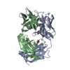
| ||||||||
| 2 | 
| ||||||||
| 単位格子 |
|
-要素
| #1: 抗体 | 分子量: 23448.883 Da / 分子数: 2 / 由来タイプ: 組換発現 / 由来: (組換発現) MUS MUSCULUS, HOMO SAPIENS / 発現宿主: unidentified (未定義) #2: 抗体 | 分子量: 23725.504 Da / 分子数: 2 / 由来タイプ: 組換発現 / 由来: (組換発現) MUS MUSCULUS, HOMO SAPIENS / 発現宿主: unidentified (未定義) #3: タンパク質・ペプチド | 分子量: 1471.771 Da / 分子数: 2 / 由来タイプ: 合成 / 由来: (合成) synthetic construct (人工物) #4: 化合物 |   分子量: 94.971 Da / 分子数: 2 / 由来タイプ: 合成 / 式: PO4 分子量: 94.971 Da / 分子数: 2 / 由来タイプ: 合成 / 式: PO4#5: 水 | ChemComp-HOH / |  分子量: 18.015 Da / 分子数: 501 / 由来タイプ: 天然 / 式: H2O 分子量: 18.015 Da / 分子数: 501 / 由来タイプ: 天然 / 式: H2O |
|---|
-実験情報
-実験
| 実験 | 手法: X線回折 / 使用した結晶の数: 1 |
|---|
- 試料調製
試料調製
| 結晶 | マシュー密度: 2.89 Å3/Da / 溶媒含有率: 57.37 % |
|---|---|
| 結晶化 | 温度: 293 K / 手法: 蒸気拡散法, ハンギングドロップ法 詳細: 0.1 M citrate, 0.1 M sodium hydrogen phosphate, 0.5 M potassium hydrogen phosphate, 1.6 M sodium dihydrogen phosphate |
-データ収集
| 回折 | 平均測定温度: 100 K |
|---|---|
| 放射光源 | 由来: 回転陽極 / タイプ: RIGAKU MICROMAX-007 HF / 波長: 1.5418 Å |
| 検出器 | タイプ: RIGAKU RAXIS IV++ / 検出器: IMAGE PLATE / 日付: 2016年1月12日 |
| 放射 | プロトコル: SINGLE WAVELENGTH / 単色(M)・ラウエ(L): M / 散乱光タイプ: x-ray |
| 放射波長 | 波長: 1.5418 Å / 相対比: 1 |
| 反射 | 解像度: 2.477→32.53 Å / Num. obs: 40620 / % possible obs: 99.1 % / 冗長度: 5.1 % / Rmerge(I) obs: 0.064 / Net I/σ(I): 22.9 |
| 反射 シェル | 解像度: 2.48→2.54 Å / 冗長度: 3.5 % / Rmerge(I) obs: 0.27 / % possible all: 88.9 |
- 解析
解析
| ソフトウェア |
| ||||||||||||||||||||||||||||||||||||||||||||||||||||||||||||||||||||||||||||||||||||||||||||||||||||||||||||||||
|---|---|---|---|---|---|---|---|---|---|---|---|---|---|---|---|---|---|---|---|---|---|---|---|---|---|---|---|---|---|---|---|---|---|---|---|---|---|---|---|---|---|---|---|---|---|---|---|---|---|---|---|---|---|---|---|---|---|---|---|---|---|---|---|---|---|---|---|---|---|---|---|---|---|---|---|---|---|---|---|---|---|---|---|---|---|---|---|---|---|---|---|---|---|---|---|---|---|---|---|---|---|---|---|---|---|---|---|---|---|---|---|---|---|
| 精密化 | 構造決定の手法: 分子置換 開始モデル: 4gw1 解像度: 2.477→32.529 Å / SU ML: 0.24 / 交差検証法: FREE R-VALUE / σ(F): 1.36 / 位相誤差: 19.47
| ||||||||||||||||||||||||||||||||||||||||||||||||||||||||||||||||||||||||||||||||||||||||||||||||||||||||||||||||
| 溶媒の処理 | 減衰半径: 0.9 Å / VDWプローブ半径: 1.11 Å | ||||||||||||||||||||||||||||||||||||||||||||||||||||||||||||||||||||||||||||||||||||||||||||||||||||||||||||||||
| 精密化ステップ | サイクル: LAST / 解像度: 2.477→32.529 Å
| ||||||||||||||||||||||||||||||||||||||||||||||||||||||||||||||||||||||||||||||||||||||||||||||||||||||||||||||||
| 拘束条件 |
| ||||||||||||||||||||||||||||||||||||||||||||||||||||||||||||||||||||||||||||||||||||||||||||||||||||||||||||||||
| LS精密化 シェル |
|
