 Movie
Movie Controller

[English] 日本語
 Yorodumi
- PDB-6azk: Structure of cetuximab with aminoheptanoic acid-linked N-(3-hydro... -
+ Open data

- Basic information
| Entry | Database: PDB / ID: 6azk | |||||||||
|---|---|---|---|---|---|---|---|---|---|---|
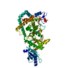
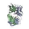
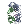
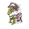
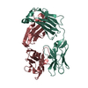
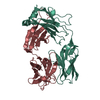
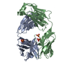
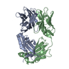
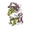
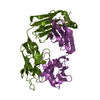
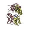
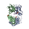
| Title | Structure of cetuximab with aminoheptanoic acid-linked N-(3-hydroxypropyl)-L-arginine meditope variant | |||||||||
 Components Components |
| |||||||||
 Keywords Keywords | IMMUNE SYSTEM / antibody / anti-EGFR | |||||||||
| Function / homology |  Function and homology information Function and homology informationCD22 mediated BCR regulation / Fc epsilon receptor (FCERI) signaling / Classical antibody-mediated complement activation / IgG immunoglobulin complex / Initial triggering of complement / immunoglobulin mediated immune response / FCGR activation / Role of LAT2/NTAL/LAB on calcium mobilization / immunoglobulin complex / Role of phospholipids in phagocytosis ...CD22 mediated BCR regulation / Fc epsilon receptor (FCERI) signaling / Classical antibody-mediated complement activation / IgG immunoglobulin complex / Initial triggering of complement / immunoglobulin mediated immune response / FCGR activation / Role of LAT2/NTAL/LAB on calcium mobilization / immunoglobulin complex / Role of phospholipids in phagocytosis / Scavenging of heme from plasma / antigen binding / FCERI mediated Ca+2 mobilization / FCGR3A-mediated IL10 synthesis / Regulation of Complement cascade / Antigen activates B Cell Receptor (BCR) leading to generation of second messengers / B cell receptor signaling pathway / Cell surface interactions at the vascular wall / FCGR3A-mediated phagocytosis / FCERI mediated MAPK activation / Regulation of actin dynamics for phagocytic cup formation / Immunoregulatory interactions between a Lymphoid and a non-Lymphoid cell / FCERI mediated NF-kB activation / blood microparticle / Potential therapeutics for SARS / adaptive immune response / immune response / : / extracellular exosome / extracellular region / plasma membrane Similarity search - Function | |||||||||
| Biological species |   Homo sapiens (human) Homo sapiens (human)synthetic construct (others) | |||||||||
| Method |  X-RAY DIFFRACTION / MOLECULAR REPLACEMENT / Resolution: 2.477 Å X-RAY DIFFRACTION / MOLECULAR REPLACEMENT / Resolution: 2.477 Å | |||||||||
 Authors Authors | Bzymek, K.P. / Williams, J.C. | |||||||||
 Citation Citation | Journal: Acta Crystallogr F Struct Biol Commun / Year: 2017 Title: Meditope-Fab interaction: threading the hole. Authors: Bzymek, K.P. / Ma, Y. / Avery, K.N. / Horne, D.A. / Williams, J.C. | |||||||||
| History |
|
- Structure visualization
| Structure viewer | Molecule: MolmilJmol/JSmol |
|---|
- Downloads & links
-Download
| PDBx/mmCIF format | 6azk.cif.gz | 194.7 KB | Display | PDBx/mmCIF format |
|---|---|---|---|---|
| PDB format | pdb6azk.ent.gz | 152.6 KB | Display | PDB format |
| PDBx/mmJSON format | 6azk.json.gz | Tree view | PDBx/mmJSON format | |
| Others |  Other downloads Other downloads |
-Validation report
| Arichive directory | https://data.pdbj.org/pub/pdb/validation_reports/az/6azkftp://data.pdbj.org/pub/pdb/validation_reports/az/6azk | HTTPS FTP |
|---|
-Related structure data
| Related structure data | 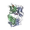 6au5C 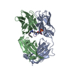 6axpC 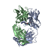 6aynC 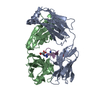 6azlC  4gw1S C: citing same article ( S: Starting model for refinement |
|---|---|
| Similar structure data |


-Links
 PDBj
PDBj


- Assembly
| Deposited unit | 
| ||||||||
|---|---|---|---|---|---|---|---|---|---|
| 1 | 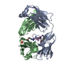
| ||||||||
| 2 | 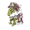
| ||||||||
| Unit cell |
|
-Components
| #1: Antibody | Mass: 23287.705 Da / Num. of mol.: 2 Source method: isolated from a genetically manipulated source Details: chimeric murine/human antibody purified from Erbitux Source: (gene. exp.) Homo sapiens (human)Production host: unidentified (others) / References: UniProt: P01834 #2: Antibody | Mass: 23725.504 Da / Num. of mol.: 2 Source method: isolated from a genetically manipulated source Details: chimeric murine/human antibody purified from Erbitux Source: (gene. exp.) Homo sapiens (human)Production host: unidentified (others) / References: UniProt: S6B291 #3: Protein/peptide | Mass: 1450.726 Da / Num. of mol.: 2 / Source method: obtained synthetically / Source: (synth.) synthetic construct (others) #4: Chemical |   Mass: 96.063 Da / Num. of mol.: 2 / Source method: obtained synthetically / Formula: SO4 Mass: 96.063 Da / Num. of mol.: 2 / Source method: obtained synthetically / Formula: SO4#5: Water | ChemComp-HOH / |  Mass: 18.015 Da / Num. of mol.: 526 / Source method: isolated from a natural source / Formula: H2O Mass: 18.015 Da / Num. of mol.: 526 / Source method: isolated from a natural source / Formula: H2OSequence details | Sequence of cetuximab Fab light chain in this entry matches to the sequence of cetuximab Fab light ...Sequence of cetuximab Fab light chain in this entry matches to the sequence of cetuximab Fab light chain from the first deposited cetuximab structure in PDB (1yy8) | |
|---|
-Experimental details
-Experiment
| Experiment | Method: X-RAY DIFFRACTION / Number of used crystals: 1 |
|---|
- Sample preparation
| Crystal | Density Matthews: 2.92 Å3/Da / Density % sol: 57.86 % |
|---|---|
| Crystal grow | Temperature: 293 K / Method: vapor diffusion, hanging drop / Details: 0.1 M citrate pH 5, 1.8 M ammonium sulfate |
-Data collection
| Diffraction | Mean temperature: 100 K |
|---|---|
| Diffraction source | Source: ROTATING ANODE / Type: RIGAKU MICROMAX-007 HF / Wavelength: 1.5418 Å |
| Detector | Type: RIGAKU RAXIS IV++ / Detector: IMAGE PLATE / Date: Apr 26, 2012 |
| Radiation | Protocol: SINGLE WAVELENGTH / Monochromatic (M) / Laue (L): M / Scattering type: x-ray |
| Radiation wavelength | Wavelength: 1.5418 Å / Relative weight: 1 |
| Reflection | Resolution: 2.477→33.113 Å / Num. obs: 40781 / % possible obs: 98.8 % / Redundancy: 4.8 % / CC1/2: 0.998 / Rmerge(I) obs: 0.067 / Net I/σ(I): 19.8 |
| Reflection shell | Resolution: 2.48→2.54 Å / Redundancy: 3.5 % / Rmerge(I) obs: 0.207 / Mean I/σ(I) obs: 6.5 / Num. unique obs: 2690 / CC1/2: 0.954 / % possible all: 90.1 |
- Processing
| Software |
| ||||||||||||||||||||||||||||||||||||||||||||||||||||||||||||||||||||||||||||||||||||||||||||||||||||||||||||||||
|---|---|---|---|---|---|---|---|---|---|---|---|---|---|---|---|---|---|---|---|---|---|---|---|---|---|---|---|---|---|---|---|---|---|---|---|---|---|---|---|---|---|---|---|---|---|---|---|---|---|---|---|---|---|---|---|---|---|---|---|---|---|---|---|---|---|---|---|---|---|---|---|---|---|---|---|---|---|---|---|---|---|---|---|---|---|---|---|---|---|---|---|---|---|---|---|---|---|---|---|---|---|---|---|---|---|---|---|---|---|---|---|---|---|
| Refinement | Method to determine structure: MOLECULAR REPLACEMENT Starting model: 4gw1 Resolution: 2.477→33.113 Å / SU ML: 0.29 / Cross valid method: FREE R-VALUE / σ(F): 2.02 / Phase error: 19.04
| ||||||||||||||||||||||||||||||||||||||||||||||||||||||||||||||||||||||||||||||||||||||||||||||||||||||||||||||||
| Solvent computation | Shrinkage radii: 0.9 Å / VDW probe radii: 1.11 Å | ||||||||||||||||||||||||||||||||||||||||||||||||||||||||||||||||||||||||||||||||||||||||||||||||||||||||||||||||
| Refinement step | Cycle: LAST / Resolution: 2.477→33.113 Å
| ||||||||||||||||||||||||||||||||||||||||||||||||||||||||||||||||||||||||||||||||||||||||||||||||||||||||||||||||
| Refine LS restraints |
| ||||||||||||||||||||||||||||||||||||||||||||||||||||||||||||||||||||||||||||||||||||||||||||||||||||||||||||||||
| LS refinement shell |
|
