 Movie
Movie Controller


+ Open data

- Basic information
| Entry | Database: PDB / ID: 5hy0 | ||||||
|---|---|---|---|---|---|---|---|
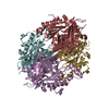
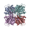
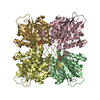
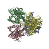
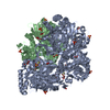
| Title | orotic acid hydrolase | ||||||
 Components Components | Ring-opening amidohydrolase | ||||||
 Keywords Keywords | HYDROLASE / Toblerone fold / pyrimidine catabolism | ||||||
| Function / homology |  Function and homology information Function and homology informationHydrolases; Acting on carbon-nitrogen bonds, other than peptide bonds; In cyclic amides / hydrolase activity, acting on carbon-nitrogen (but not peptide) bonds, in cyclic amides Similarity search - Function | ||||||
| Biological species |  Frankia sp. Eul1b (bacteria) Frankia sp. Eul1b (bacteria) | ||||||
| Method |  X-RAY DIFFRACTION / SYNCHROTRON / MOLECULAR REPLACEMENT / Resolution: 2.4 Å X-RAY DIFFRACTION / SYNCHROTRON / MOLECULAR REPLACEMENT / Resolution: 2.4 Å | ||||||
 Authors Authors | Peat, T.S. / Balotra, S. / Wilding, M. / Newman, J. / Scott, C. | ||||||
 Citation Citation | Journal: Appl. Environ. Microbiol. / Year: 2017 Title: High-Resolution X-Ray Structures of Two Functionally Distinct Members of the Cyclic Amide Hydrolase Family of Toblerone Fold Enzymes. Authors: Peat, T.S. / Balotra, S. / Wilding, M. / Hartley, C.J. / Newman, J. / Scott, C. | ||||||
| History |
|
- Structure visualization
| Structure viewer | Molecule: MolmilJmol/JSmol |
|---|
- Downloads & links
-Download
| PDBx/mmCIF format | 5hy0.cif.gz | 274.9 KB | Display | PDBx/mmCIF format |
|---|---|---|---|---|
| PDB format | pdb5hy0.ent.gz | 218.4 KB | Display | PDB format |
| PDBx/mmJSON format | 5hy0.json.gz | Tree view | PDBx/mmJSON format | |
| Others |  Other downloads Other downloads |
-Validation report
| Arichive directory | https://data.pdbj.org/pub/pdb/validation_reports/hy/5hy0ftp://data.pdbj.org/pub/pdb/validation_reports/hy/5hy0 | HTTPS FTP |
|---|
-Related structure data
| Related structure data |  5hweC  5hxuC  5hxzC  5hy1C  5hy2C  5hy4C 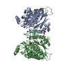 4bvqS C: citing same article ( S: Starting model for refinement |
|---|---|
| Similar structure data |


-Links
 PDBj
PDBj- Assembly
| Deposited unit | 
| ||||||||||||||||||||||||||||||||||||||||||||||||||||||||||||||||||||||||||||||||||||||||||||||||||||||||||||||||||||||||||||||||||||||||||||||||||||||
|---|---|---|---|---|---|---|---|---|---|---|---|---|---|---|---|---|---|---|---|---|---|---|---|---|---|---|---|---|---|---|---|---|---|---|---|---|---|---|---|---|---|---|---|---|---|---|---|---|---|---|---|---|---|---|---|---|---|---|---|---|---|---|---|---|---|---|---|---|---|---|---|---|---|---|---|---|---|---|---|---|---|---|---|---|---|---|---|---|---|---|---|---|---|---|---|---|---|---|---|---|---|---|---|---|---|---|---|---|---|---|---|---|---|---|---|---|---|---|---|---|---|---|---|---|---|---|---|---|---|---|---|---|---|---|---|---|---|---|---|---|---|---|---|---|---|---|---|---|---|---|---|
| 1 |
| ||||||||||||||||||||||||||||||||||||||||||||||||||||||||||||||||||||||||||||||||||||||||||||||||||||||||||||||||||||||||||||||||||||||||||||||||||||||
| Unit cell |
| ||||||||||||||||||||||||||||||||||||||||||||||||||||||||||||||||||||||||||||||||||||||||||||||||||||||||||||||||||||||||||||||||||||||||||||||||||||||
| Noncrystallographic symmetry (NCS) | NCS domain:
NCS domain segments: Component-ID: _ / Beg auth comp-ID: ARG / Beg label comp-ID: ARG / Refine code: _
NCS ensembles :
|
-Components
| #1: Protein | Mass: 42103.465 Da / Num. of mol.: 4 Source method: isolated from a genetically manipulated source Source: (gene. exp.) Frankia sp. Eul1b (bacteria) / Plasmid: pET14b variant / Production host: #2: Chemical |   Mass: 92.094 Da / Num. of mol.: 2 / Source method: obtained synthetically / Formula: C3H8O3 Mass: 92.094 Da / Num. of mol.: 2 / Source method: obtained synthetically / Formula: C3H8O3#3: Water | ChemComp-HOH / |  Mass: 18.015 Da / Num. of mol.: 350 / Source method: isolated from a natural source / Formula: H2O Mass: 18.015 Da / Num. of mol.: 350 / Source method: isolated from a natural source / Formula: H2OHas protein modification | Y | |
|---|
-Experimental details
-Experiment
| Experiment | Method: X-RAY DIFFRACTION / Number of used crystals: 1 |
|---|
- Sample preparation
| Crystal | Density Matthews: 2.6 Å3/Da / Density % sol: 52.78 % |
|---|---|
| Crystal grow | Temperature: 281 K / Method: vapor diffusion, sitting drop / pH: 7.5 Details: Protein was concentrated to 2.1 mg/mL in 100 mM NaCl and 50 mM HEPES pH 7.5; reservoir was 20% PEG 3350, 0.2 M magnesium acetate, 20 mM HEPES pH 6.8 with the Silver Bullets Bio C1 condition ...Details: Protein was concentrated to 2.1 mg/mL in 100 mM NaCl and 50 mM HEPES pH 7.5; reservoir was 20% PEG 3350, 0.2 M magnesium acetate, 20 mM HEPES pH 6.8 with the Silver Bullets Bio C1 condition of additives; 200 nL of protein added to 90 nL of reservoir and 10 nL of microseeds. PH range: 6.8 - 7.5 |
-Data collection
| Diffraction | Mean temperature: 100 K |
|---|---|
| Diffraction source | Source: SYNCHROTRON / Site: Australian Synchrotron  / Beamline: MX2 / Wavelength: 0.9537 Å / Beamline: MX2 / Wavelength: 0.9537 Å |
| Detector | Type: ADSC QUANTUM 315r / Detector: CCD / Date: Dec 6, 2014 |
| Radiation | Protocol: SINGLE WAVELENGTH / Monochromatic (M) / Laue (L): M / Scattering type: x-ray |
| Radiation wavelength | Wavelength: 0.9537 Å / Relative weight: 1 |
| Reflection | Resolution: 2.4→46.3 Å / Num. obs: 65127 / % possible obs: 97.5 % / Redundancy: 3.9 % / Net I/σ(I): 24 |
| Reflection shell | Resolution: 2.4→2.46 Å / Redundancy: 3.8 % / Mean I/σ(I) obs: 3.8 / % possible all: 83.1 |
- Processing
| Software |
| ||||||||||||||||||||||||||||||||||||||||||||||||||||||||||||||||||||||||||||||||||||||||||||||||||||||||||||||||||||||||||||||||||||||||||||||||||||||||||||||||||||||||||||||||||||||
|---|---|---|---|---|---|---|---|---|---|---|---|---|---|---|---|---|---|---|---|---|---|---|---|---|---|---|---|---|---|---|---|---|---|---|---|---|---|---|---|---|---|---|---|---|---|---|---|---|---|---|---|---|---|---|---|---|---|---|---|---|---|---|---|---|---|---|---|---|---|---|---|---|---|---|---|---|---|---|---|---|---|---|---|---|---|---|---|---|---|---|---|---|---|---|---|---|---|---|---|---|---|---|---|---|---|---|---|---|---|---|---|---|---|---|---|---|---|---|---|---|---|---|---|---|---|---|---|---|---|---|---|---|---|---|---|---|---|---|---|---|---|---|---|---|---|---|---|---|---|---|---|---|---|---|---|---|---|---|---|---|---|---|---|---|---|---|---|---|---|---|---|---|---|---|---|---|---|---|---|---|---|---|---|
| Refinement | Method to determine structure: MOLECULAR REPLACEMENT Starting model: 4BVQ Resolution: 2.4→41.5 Å / Cor.coef. Fo:Fc: 0.936 / Cor.coef. Fo:Fc free: 0.904 / SU B: 8.021 / SU ML: 0.176 / Cross valid method: THROUGHOUT / ESU R: 0.365 / ESU R Free: 0.244 / Details: HYDROGENS HAVE BEEN ADDED IN THE RIDING POSITIONS
| ||||||||||||||||||||||||||||||||||||||||||||||||||||||||||||||||||||||||||||||||||||||||||||||||||||||||||||||||||||||||||||||||||||||||||||||||||||||||||||||||||||||||||||||||||||||
| Solvent computation | Ion probe radii: 0.8 Å / Shrinkage radii: 0.8 Å / VDW probe radii: 1.2 Å | ||||||||||||||||||||||||||||||||||||||||||||||||||||||||||||||||||||||||||||||||||||||||||||||||||||||||||||||||||||||||||||||||||||||||||||||||||||||||||||||||||||||||||||||||||||||
| Displacement parameters | Biso mean: 26.693 Å2
| ||||||||||||||||||||||||||||||||||||||||||||||||||||||||||||||||||||||||||||||||||||||||||||||||||||||||||||||||||||||||||||||||||||||||||||||||||||||||||||||||||||||||||||||||||||||
| Refinement step | Cycle: 1 / Resolution: 2.4→41.5 Å
| ||||||||||||||||||||||||||||||||||||||||||||||||||||||||||||||||||||||||||||||||||||||||||||||||||||||||||||||||||||||||||||||||||||||||||||||||||||||||||||||||||||||||||||||||||||||
| Refine LS restraints |
|