 Movie
Movie Controller

[English] 日本語
 Yorodumi
- PDB-5hxu: Structure-function analysis of functionally diverse members of th... -
+ Open data

- Basic information
| Entry | Database: PDB / ID: 5hxu | ||||||
|---|---|---|---|---|---|---|---|
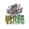
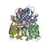
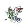
| Title | Structure-function analysis of functionally diverse members of the cyclic amide hydrolase family of Toblerone fold enzymes | ||||||
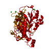
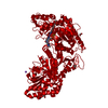
 Components Components | Barbiturase | ||||||
 Keywords Keywords | HYDROLASE / Toblerone fold / pyrimidine catabolism | ||||||
| Function / homology |  Function and homology information Function and homology informationbarbiturase / barbiturase activity / uracil catabolic process / metal ion binding Similarity search - Function | ||||||
| Biological species |  Rhodococcus erythropolis (bacteria) Rhodococcus erythropolis (bacteria) | ||||||
| Method |  X-RAY DIFFRACTION / SYNCHROTRON / MOLECULAR REPLACEMENT / Resolution: 1.83 Å X-RAY DIFFRACTION / SYNCHROTRON / MOLECULAR REPLACEMENT / Resolution: 1.83 Å | ||||||
 Authors Authors | Peat, T.S. / Balotra, S. / Wilding, M. / Newman, J. / Scott, C. | ||||||
 Citation Citation | Journal: Appl. Environ. Microbiol. / Year: 2017 Title: High-Resolution X-Ray Structures of Two Functionally Distinct Members of the Cyclic Amide Hydrolase Family of Toblerone Fold Enzymes. Authors: Peat, T.S. / Balotra, S. / Wilding, M. / Hartley, C.J. / Newman, J. / Scott, C. | ||||||
| History |
|
- Structure visualization
| Structure viewer | Molecule: MolmilJmol/JSmol |
|---|
- Downloads & links
-Download
| PDBx/mmCIF format | 5hxu.cif.gz | 167.3 KB | Display | PDBx/mmCIF format |
|---|---|---|---|---|
| PDB format | pdb5hxu.ent.gz | 129 KB | Display | PDB format |
| PDBx/mmJSON format | 5hxu.json.gz | Tree view | PDBx/mmJSON format | |
| Others |  Other downloads Other downloads |
-Validation report
| Arichive directory | https://data.pdbj.org/pub/pdb/validation_reports/hx/5hxuftp://data.pdbj.org/pub/pdb/validation_reports/hx/5hxu | HTTPS FTP |
|---|
-Related structure data
| Related structure data |  5hweC  5hxzC  5hy0C  5hy1C  5hy2C  5hy4C 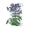 4bvqS C: citing same article ( S: Starting model for refinement |
|---|---|
| Similar structure data |


-Links
 PDBj
PDBj- Assembly
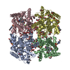
| Deposited unit | 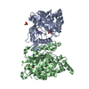
| ||||||||||||||||||
|---|---|---|---|---|---|---|---|---|---|---|---|---|---|---|---|---|---|---|---|
| 1 | 
| ||||||||||||||||||
| Unit cell |
| ||||||||||||||||||
| Components on special symmetry positions |
| ||||||||||||||||||
| Noncrystallographic symmetry (NCS) | NCS domain:
NCS domain segments: Component-ID: _ / Ens-ID: 1 / Beg auth comp-ID: GLU / Beg label comp-ID: GLU / End auth comp-ID: LEU / End label comp-ID: LEU / Refine code: _ / Auth seq-ID: 3 - 367 / Label seq-ID: 23 - 387
|
-Components
-Protein , 1 types, 2 molecules AB
| #1: Protein | Mass: 41188.172 Da / Num. of mol.: 2 Source method: isolated from a genetically manipulated source Source: (gene. exp.) Rhodococcus erythropolis (bacteria) / Gene: bar / Plasmid: pET14b variant / Production host: |
|---|
-Non-polymers , 5 types, 626 molecules 


| #2: Chemical |  Mass: 96.063 Da / Num. of mol.: 3 / Source method: obtained synthetically / Formula: SO4 Mass: 96.063 Da / Num. of mol.: 3 / Source method: obtained synthetically / Formula: SO4#3: Chemical |  Mass: 190.154 Da / Num. of mol.: 2 / Source method: obtained synthetically / Formula: C6H10N2O5 / Comment: pH buffer*YM Mass: 190.154 Da / Num. of mol.: 2 / Source method: obtained synthetically / Formula: C6H10N2O5 / Comment: pH buffer*YM#4: Chemical |  Mass: 22.990 Da / Num. of mol.: 2 / Source method: obtained synthetically / Formula: Na Mass: 22.990 Da / Num. of mol.: 2 / Source method: obtained synthetically / Formula: Na#5: Chemical |  Mass: 35.453 Da / Num. of mol.: 2 / Source method: obtained synthetically / Formula: Cl Mass: 35.453 Da / Num. of mol.: 2 / Source method: obtained synthetically / Formula: Cl#6: Water | ChemComp-HOH / | Mass: 18.015 Da / Num. of mol.: 617 / Source method: isolated from a natural source / Formula: H2O |
|---|
-Experimental details
-Experiment
| Experiment | Method: X-RAY DIFFRACTION / Number of used crystals: 1 |
|---|
- Sample preparation
| Crystal | Density Matthews: 2.43 Å3/Da / Density % sol: 49.5 % |
|---|---|
| Crystal grow | Temperature: 293 K / Method: vapor diffusion, sitting drop / pH: 9 Details: Protein at 15 mg/mL; reservoir was 2.5 M ammonium sulfate with 10% (v/v) MMT buffer at pH 9; sitting drop setup with 150 nL plus 150 nL drops; crystals cryo-protected with AP/E core 150 ...Details: Protein at 15 mg/mL; reservoir was 2.5 M ammonium sulfate with 10% (v/v) MMT buffer at pH 9; sitting drop setup with 150 nL plus 150 nL drops; crystals cryo-protected with AP/E core 150 basestock (Mobile 1, Australia). |
-Data collection
| Diffraction | Mean temperature: 100 K |
|---|---|
| Diffraction source | Source: SYNCHROTRON / Site: Australian Synchrotron  / Beamline: MX2 / Wavelength: 1.28221 Å / Beamline: MX2 / Wavelength: 1.28221 Å |
| Detector | Type: ADSC QUANTUM 315r / Detector: CCD / Date: Feb 18, 2014 |
| Radiation | Protocol: SINGLE WAVELENGTH / Monochromatic (M) / Laue (L): M / Scattering type: x-ray |
| Radiation wavelength | Wavelength: 1.28221 Å / Relative weight: 1 |
| Reflection | Resolution: 1.83→45.7 Å / Num. obs: 67015 / % possible obs: 98.1 % / Redundancy: 26.6 % / Net I/σ(I): 22.2 |
| Reflection shell | Resolution: 1.83→1.87 Å / Redundancy: 20.5 % / Mean I/σ(I) obs: 3.5 / % possible all: 81 |
- Processing
| Software |
| ||||||||||||||||||||||||||||||||||||||||||||||||||||||||||||||||||||||||||||||||||||||||||||||||||||||||||||||||||||||||||||||||||||||||||||||||||||||||||||||||||||||||||||||||||||||
|---|---|---|---|---|---|---|---|---|---|---|---|---|---|---|---|---|---|---|---|---|---|---|---|---|---|---|---|---|---|---|---|---|---|---|---|---|---|---|---|---|---|---|---|---|---|---|---|---|---|---|---|---|---|---|---|---|---|---|---|---|---|---|---|---|---|---|---|---|---|---|---|---|---|---|---|---|---|---|---|---|---|---|---|---|---|---|---|---|---|---|---|---|---|---|---|---|---|---|---|---|---|---|---|---|---|---|---|---|---|---|---|---|---|---|---|---|---|---|---|---|---|---|---|---|---|---|---|---|---|---|---|---|---|---|---|---|---|---|---|---|---|---|---|---|---|---|---|---|---|---|---|---|---|---|---|---|---|---|---|---|---|---|---|---|---|---|---|---|---|---|---|---|---|---|---|---|---|---|---|---|---|---|---|
| Refinement | Method to determine structure: MOLECULAR REPLACEMENT Starting model: 4bvq Resolution: 1.83→41.5 Å / Cor.coef. Fo:Fc: 0.913 / Cor.coef. Fo:Fc free: 0.89 / SU B: 3.122 / SU ML: 0.095 / Cross valid method: THROUGHOUT / ESU R: 0.16 / ESU R Free: 0.149 / Details: HYDROGENS HAVE BEEN ADDED IN THE RIDING POSITIONS
| ||||||||||||||||||||||||||||||||||||||||||||||||||||||||||||||||||||||||||||||||||||||||||||||||||||||||||||||||||||||||||||||||||||||||||||||||||||||||||||||||||||||||||||||||||||||
| Solvent computation | Ion probe radii: 0.8 Å / Shrinkage radii: 0.8 Å / VDW probe radii: 1.2 Å | ||||||||||||||||||||||||||||||||||||||||||||||||||||||||||||||||||||||||||||||||||||||||||||||||||||||||||||||||||||||||||||||||||||||||||||||||||||||||||||||||||||||||||||||||||||||
| Displacement parameters | Biso mean: 25.162 Å2
| ||||||||||||||||||||||||||||||||||||||||||||||||||||||||||||||||||||||||||||||||||||||||||||||||||||||||||||||||||||||||||||||||||||||||||||||||||||||||||||||||||||||||||||||||||||||
| Refinement step | Cycle: 1 / Resolution: 1.83→41.5 Å
| ||||||||||||||||||||||||||||||||||||||||||||||||||||||||||||||||||||||||||||||||||||||||||||||||||||||||||||||||||||||||||||||||||||||||||||||||||||||||||||||||||||||||||||||||||||||
| Refine LS restraints |
|