 Movie
Movie Controller


+ Open data

- Basic information
| Entry | Database: PDB / ID: 5hwe | ||||||
|---|---|---|---|---|---|---|---|
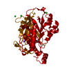
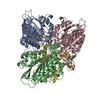
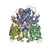
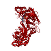
| Title | high resolution structure of barbiturase | ||||||
 Components Components | Barbiturase | ||||||
 Keywords Keywords | HYDROLASE / Toblerone fold / pyrimidine catabolism | ||||||
| Function / homology |  Function and homology information Function and homology informationbarbiturase / barbiturase activity / uracil catabolic process / metal ion binding Similarity search - Function | ||||||
| Biological species |  Rhodococcus erythropolis (bacteria) Rhodococcus erythropolis (bacteria) | ||||||
| Method |  X-RAY DIFFRACTION / SYNCHROTRON / MOLECULAR REPLACEMENT / Resolution: 1.71 Å X-RAY DIFFRACTION / SYNCHROTRON / MOLECULAR REPLACEMENT / Resolution: 1.71 Å | ||||||
 Authors Authors | Peat, T.S. / Scott, C. | ||||||
 Citation Citation | Journal: Appl. Environ. Microbiol. / Year: 2017 Title: High-Resolution X-Ray Structures of Two Functionally Distinct Members of the Cyclic Amide Hydrolase Family of Toblerone Fold Enzymes. Authors: Peat, T.S. / Balotra, S. / Wilding, M. / Hartley, C.J. / Newman, J. / Scott, C. | ||||||
| History |
|
- Structure visualization
| Structure viewer | Molecule: MolmilJmol/JSmol |
|---|
- Downloads & links
-Download
| PDBx/mmCIF format | 5hwe.cif.gz | 94.2 KB | Display | PDBx/mmCIF format |
|---|---|---|---|---|
| PDB format | pdb5hwe.ent.gz | 68.3 KB | Display | PDB format |
| PDBx/mmJSON format | 5hwe.json.gz | Tree view | PDBx/mmJSON format | |
| Others |  Other downloads Other downloads |
-Validation report
| Arichive directory | https://data.pdbj.org/pub/pdb/validation_reports/hw/5hweftp://data.pdbj.org/pub/pdb/validation_reports/hw/5hwe | HTTPS FTP |
|---|
-Related structure data
| Related structure data |  5hxuC  5hxzC  5hy0C  5hy1C  5hy2C  5hy4C 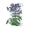 4bvqS C: citing same article ( S: Starting model for refinement |
|---|---|
| Similar structure data |


-Links
 PDBj
PDBj- Assembly
| Deposited unit | 
| |||||||||
|---|---|---|---|---|---|---|---|---|---|---|
| 1 | 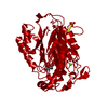
| |||||||||
| Unit cell |
| |||||||||
| Components on special symmetry positions |
|
-Components
| #1: Protein | Mass: 41188.172 Da / Num. of mol.: 1 Source method: isolated from a genetically manipulated source Source: (gene. exp.) Rhodococcus erythropolis (bacteria) / Gene: bar / Plasmid: pET14b variant / Production host: |
|---|---|
| #2: Chemical | ChemComp-MG /   Mass: 24.305 Da / Num. of mol.: 1 / Source method: obtained synthetically / Formula: Mg Mass: 24.305 Da / Num. of mol.: 1 / Source method: obtained synthetically / Formula: Mg |
| #3: Chemical | ChemComp-SO4 /   Mass: 96.063 Da / Num. of mol.: 1 / Source method: obtained synthetically / Formula: SO4 Mass: 96.063 Da / Num. of mol.: 1 / Source method: obtained synthetically / Formula: SO4 |
| #4: Chemical | ChemComp-MHA / (  Mass: 190.154 Da / Num. of mol.: 1 / Source method: obtained synthetically / Formula: C6H10N2O5 / Comment: pH buffer*YM Mass: 190.154 Da / Num. of mol.: 1 / Source method: obtained synthetically / Formula: C6H10N2O5 / Comment: pH buffer*YM |
| #5: Water | ChemComp-HOH /  Mass: 18.015 Da / Num. of mol.: 295 / Source method: isolated from a natural source / Formula: H2O Mass: 18.015 Da / Num. of mol.: 295 / Source method: isolated from a natural source / Formula: H2O |
-Experimental details
-Experiment
| Experiment | Method: X-RAY DIFFRACTION / Number of used crystals: 1 |
|---|
- Sample preparation
| Crystal | Density Matthews: 2.13 Å3/Da / Density % sol: 42.15 % |
|---|---|
| Crystal grow | Temperature: 293 K / Method: vapor diffusion, sitting drop / pH: 6.5 Details: Protein at 16 mg/,mL in 50 mM ADA pH 6.5, 50 mM NaCl; reservoir of 2.45 M ammonium sulfate, 10% glycerol |
-Data collection
| Diffraction | Mean temperature: 100 K |
|---|---|
| Diffraction source | Source: SYNCHROTRON / Site: Australian Synchrotron  / Beamline: MX2 / Wavelength: 0.9537 Å / Beamline: MX2 / Wavelength: 0.9537 Å |
| Detector | Type: ADSC QUANTUM 315r / Detector: CCD / Date: Mar 21, 2014 |
| Radiation | Protocol: SINGLE WAVELENGTH / Monochromatic (M) / Laue (L): M / Scattering type: x-ray |
| Radiation wavelength | Wavelength: 0.9537 Å / Relative weight: 1 |
| Reflection | Resolution: 1.707→41.5 Å / Num. obs: 36498 / % possible obs: 99.7 % / Redundancy: 7.2 % / Net I/σ(I): 13.2 |
| Reflection shell | Resolution: 1.707→1.74 Å / Redundancy: 6.4 % / Mean I/σ(I) obs: 2.3 / % possible all: 93.8 |
- Processing
| Software |
| ||||||||||||||||||||||||||||||||||||||||||||||||||||||||||||||||||||||||||||||||||||||||||||||||||||||||||||||||||||||||||||||||||||||||||||||||||||||||||||||||||||||||||||||||||||||
|---|---|---|---|---|---|---|---|---|---|---|---|---|---|---|---|---|---|---|---|---|---|---|---|---|---|---|---|---|---|---|---|---|---|---|---|---|---|---|---|---|---|---|---|---|---|---|---|---|---|---|---|---|---|---|---|---|---|---|---|---|---|---|---|---|---|---|---|---|---|---|---|---|---|---|---|---|---|---|---|---|---|---|---|---|---|---|---|---|---|---|---|---|---|---|---|---|---|---|---|---|---|---|---|---|---|---|---|---|---|---|---|---|---|---|---|---|---|---|---|---|---|---|---|---|---|---|---|---|---|---|---|---|---|---|---|---|---|---|---|---|---|---|---|---|---|---|---|---|---|---|---|---|---|---|---|---|---|---|---|---|---|---|---|---|---|---|---|---|---|---|---|---|---|---|---|---|---|---|---|---|---|---|---|
| Refinement | Method to determine structure: MOLECULAR REPLACEMENT Starting model: 4BVQ Resolution: 1.71→41.5 Å / Cor.coef. Fo:Fc: 0.97 / Cor.coef. Fo:Fc free: 0.949 / SU B: 2.17 / SU ML: 0.07 / Cross valid method: THROUGHOUT / ESU R: 0.098 / ESU R Free: 0.101 / Details: HYDROGENS HAVE BEEN ADDED IN THE RIDING POSITIONS
| ||||||||||||||||||||||||||||||||||||||||||||||||||||||||||||||||||||||||||||||||||||||||||||||||||||||||||||||||||||||||||||||||||||||||||||||||||||||||||||||||||||||||||||||||||||||
| Solvent computation | Ion probe radii: 0.8 Å / Shrinkage radii: 0.8 Å / VDW probe radii: 1.2 Å | ||||||||||||||||||||||||||||||||||||||||||||||||||||||||||||||||||||||||||||||||||||||||||||||||||||||||||||||||||||||||||||||||||||||||||||||||||||||||||||||||||||||||||||||||||||||
| Displacement parameters | Biso mean: 19.557 Å2
| ||||||||||||||||||||||||||||||||||||||||||||||||||||||||||||||||||||||||||||||||||||||||||||||||||||||||||||||||||||||||||||||||||||||||||||||||||||||||||||||||||||||||||||||||||||||
| Refinement step | Cycle: LAST / Resolution: 1.71→41.5 Å
| ||||||||||||||||||||||||||||||||||||||||||||||||||||||||||||||||||||||||||||||||||||||||||||||||||||||||||||||||||||||||||||||||||||||||||||||||||||||||||||||||||||||||||||||||||||||
| Refine LS restraints |
|