- PDB-3mbh: Crystal structure of a putative phosphomethylpyrimidine kinase (B... -
+
Open data
ID or keywords:
Loading...
-
Basic information
Entry
Database: PDB / ID: 3mbh
Title
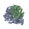
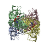
Crystal structure of a putative phosphomethylpyrimidine kinase (BT_4458) from BACTEROIDES THETAIOTAOMICRON VPI-5482 at 2.00 A resolution (orthorhombic form with pyridoxal)
Components
Putative phosphomethylpyrimidine kinase
Keywords
TRANSFERASE / Structural Genomics / Joint Center for Structural Genomics / JCSG / Protein Structure Initiative / PSI-2 / Kinase
Function / homology
Function and homology information
pyridoxal 5'-phosphate salvage / pyridoxal kinase / pyridoxal kinase activity / ATP binding / metal ion binding / cytosol Similarity search - Function
Mass: 18.015 Da / Num. of mol.: 1022 / Source method: isolated from a natural source / Formula: H2O
Has protein modification
Y
Sequence details
THE CONSTRUCT WAS EXPRESSED WITH A PURIFICATION TAG MGSDKIHHHHHHENLYFQG. THE TAG WAS REMOVED WITH ...THE CONSTRUCT WAS EXPRESSED WITH A PURIFICATION TAG MGSDKIHHHHHHENLYFQG. THE TAG WAS REMOVED WITH TEV PROTEASE LEAVING ONLY A GLYCINE (0) FOLLOWED BY THE TARGET SEQUENCE.
-
Experimental details
-
Experiment
Experiment
Method: X-RAY DIFFRACTION / Number of used crystals: 1
-
Sample preparation
Crystal
Density Matthews: 2.32 Å3/Da / Density % sol: 46.98 %
Crystal grow
Temperature: 277 K / Method: vapor diffusion, sitting drop / pH: 7.5 Details: 20.00% polyethylene glycol 3000, 0.20M sodium chloride, 0.1M HEPES pH 7.5, Additive: 0.001 M pyridoxal, NANODROP, VAPOR DIFFUSION, SITTING DROP, temperature 277K
Monochromator: Double crystal monochromator / Protocol: SINGLE WAVELENGTH / Monochromatic (M) / Laue (L): M / Scattering type: x-ray
Radiation wavelength
Wavelength: 0.97911 Å / Relative weight: 1
Reflection
Resolution: 2→29.814 Å / Num. obs: 125843 / % possible obs: 98.3 % / Observed criterion σ(I): -3 / Biso Wilson estimate: 30.527 Å2 / Rmerge(I) obs: 0.048 / Net I/σ(I): 12.12
Reflection shell
Resolution (Å)
Rmerge(I) obs
Mean I/σ(I) obs
Num. measured obs
Num. unique obs
Diffraction-ID
% possible all
2-2.07
0.554
1.5
44417
23259
1
97.5
2.07-2.15
0.373
2.3
44718
23311
1
98.7
2.15-2.25
0.283
3.1
46721
24394
1
97.6
2.25-2.37
0.205
4.1
46678
24312
1
98.4
2.37-2.52
0.152
5.4
47066
24419
1
99.1
2.52-2.71
0.103
7.8
45701
23660
1
98.9
2.71-2.99
0.066
11.6
47997
24763
1
99
2.99-3.42
0.038
18.6
46384
23878
1
98.7
3.42-4.3
0.022
29.8
45999
23659
1
97.7
4.3-29.814
0.018
37.1
46748
23956
1
97.5
-
Phasing
Phasing
Method: SAD
-
Processing
Software
Name
Version
Classification
NB
REFMAC
5.5.0053
refinement
PHENIX
refinement
SHELX
phasing
MolProbity
3beta29
modelbuilding
XSCALE
datascaling
PDB_EXTRACT
3.006
dataextraction
XDS
datareduction
SHELXD
phasing
autoSHARP
phasing
Refinement
Method to determine structure: SAD / Resolution: 2→29.814 Å / Cor.coef. Fo:Fc: 0.965 / Cor.coef. Fo:Fc free: 0.948 / Occupancy max: 1 / Occupancy min: 0.15 / SU B: 8.244 / SU ML: 0.103 / TLS residual ADP flag: LIKELY RESIDUAL / Cross valid method: THROUGHOUT / σ(F): 0 / ESU R: 0.174 / ESU R Free: 0.15 Stereochemistry target values: MAXIMUM LIKELIHOOD WITH PHASES Details: 1. HYDROGENS HAVE BEEN ADDED IN THE RIDING POSITIONS. 2. ATOM RECORD CONTAINS RESIDUAL B FACTORS ONLY. 3. A MET-INHIBITION PROTOCOL WAS USED FOR SELENOMETHIONINE INCORPORATION DURING PROTEIN ...Details: 1. HYDROGENS HAVE BEEN ADDED IN THE RIDING POSITIONS. 2. ATOM RECORD CONTAINS RESIDUAL B FACTORS ONLY. 3. A MET-INHIBITION PROTOCOL WAS USED FOR SELENOMETHIONINE INCORPORATION DURING PROTEIN EXPRESSION. THE OCCUPANCY OF THE SE ATOMS IN THE MSE RESIDUES WAS REDUCED TO 0.75 TO ACCOUNT FOR THE REDUCED SCATTERING POWER DUE TO PARTIAL S-MET INCORPORATION. 4. PYRIDOXAL (PXL) MOLECULES FROM THE CRYSTALLIZATION DROP ADDITIVE AND CHLORIDE ANIONS (CL) FROM CRYSTALLIZATION CONDITIONS ARE MODELED IN THE STRUCTURE.
Rfactor
Num. reflection
% reflection
Selection details
Rfree
0.206
6306
5 %
RANDOM
Rwork
0.167
-
-
-
obs
0.169
125765
99.57 %
-
Solvent computation
Ion probe radii: 0.8 Å / Shrinkage radii: 0.8 Å / VDW probe radii: 1.2 Å / Solvent model: MASK
In the structure databanks used in Yorodumi, some data are registered as the other names, "COVID-19 virus" and "2019-nCoV". Here are the details of the virus and the list of structure data.
Jan 31, 2019. EMDB accession codes are about to change! (news from PDBe EMDB page)
EMDB accession codes are about to change! (news from PDBe EMDB page)
The allocation of 4 digits for EMDB accession codes will soon come to an end. Whilst these codes will remain in use, new EMDB accession codes will include an additional digit and will expand incrementally as the available range of codes is exhausted. The current 4-digit format prefixed with “EMD-” (i.e. EMD-XXXX) will advance to a 5-digit format (i.e. EMD-XXXXX), and so on. It is currently estimated that the 4-digit codes will be depleted around Spring 2019, at which point the 5-digit format will come into force.
The EM Navigator/Yorodumi systems omit the EMD- prefix.
Related info.:Q: What is EMD? / ID/Accession-code notation in Yorodumi/EM Navigator
Yorodumi is a browser for structure data from EMDB, PDB, SASBDB, etc.
This page is also the successor to EM Navigator detail page, and also detail information page/front-end page for Omokage search.
The word "yorodu" (or yorozu) is an old Japanese word meaning "ten thousand". "mi" (miru) is to see.
Related info.:EMDB / PDB / SASBDB / Comparison of 3 databanks / Yorodumi Search / Aug 31, 2016. New EM Navigator & Yorodumi / Yorodumi Papers / Jmol/JSmol / Function and homology information / Changes in new EM Navigator and Yorodumi
 Movie
Movie Controller

 Yorodumi
 Open data
 Basic information
 Components
 Keywords
 Function and homology information
 Bacteroides thetaiotaomicron (bacteria)
 X-RAY DIFFRACTION /
  Authors
 Citation
 Structure visualization
 Downloads & links
 Other downloads


 PDBj
PDBj Assembly
 Mass: 167.162 Da / Num. of mol.: 6 / Source method: obtained synthetically / Formula: C8H9NO3
 Mass: 35.453 Da / Num. of mol.: 4 / Source method: obtained synthetically / Formula: Cl
 Mass: 18.015 Da / Num. of mol.: 1022 / Source method: isolated from a natural source / Formula: H2O
 Sample preparation
 / Beamline: BL9-2 / Wavelength: 0.97911
  Processing