 Movie
Movie Controller

[English] 日本語
 Yorodumi
- PDB-5hmc: Crystal structure of S. sahachiroi AziG complexed with 5-methyl n... -
+ Open data

- Basic information
| Entry | Database: PDB / ID: 5hmc | ||||||
|---|---|---|---|---|---|---|---|
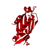
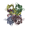
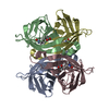
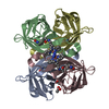
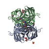
| Title | Crystal structure of S. sahachiroi AziG complexed with 5-methyl naphthoic acid | ||||||
 Components Components | Azi13 | ||||||
 Keywords Keywords | HYDROLASE / azinomycin biosynthesis / polyketide synthase / thioesterase / naphthoate | ||||||
| Function / homology |  Function and homology information Function and homology information | ||||||
| Biological species |  Streptomyces sahachiroi (bacteria) Streptomyces sahachiroi (bacteria) | ||||||
| Method |  X-RAY DIFFRACTION / SYNCHROTRON / FOURIER SYNTHESIS / Resolution: 2.2 Å X-RAY DIFFRACTION / SYNCHROTRON / FOURIER SYNTHESIS / Resolution: 2.2 Å | ||||||
 Authors Authors | Zhang, Y. / Erb, M.S. / Ealick, S.E. | ||||||
 Citation Citation | Journal: Biochemistry / Year: 2016 Title: Polyketide Ring Expansion Mediated by a Thioesterase, Chain Elongation and Cyclization Domain, in Azinomycin Biosynthesis: Characterization of AziB and AziG. Authors: Mori, S. / Simkhada, D. / Zhang, H. / Erb, M.S. / Zhang, Y. / Williams, H. / Fedoseyenko, D. / Russell, W.K. / Kim, D. / Fleer, N. / Ealick, S.E. / Watanabe, C.M. | ||||||
| History |
|
- Structure visualization
| Structure viewer | Molecule: MolmilJmol/JSmol |
|---|
- Downloads & links
-Download
| PDBx/mmCIF format | 5hmc.cif.gz | 39.1 KB | Display | PDBx/mmCIF format |
|---|---|---|---|---|
| PDB format | pdb5hmc.ent.gz | 25.5 KB | Display | PDB format |
| PDBx/mmJSON format | 5hmc.json.gz | Tree view | PDBx/mmJSON format | |
| Others |  Other downloads Other downloads |
-Validation report
| Arichive directory | https://data.pdbj.org/pub/pdb/validation_reports/hm/5hmcftp://data.pdbj.org/pub/pdb/validation_reports/hm/5hmc | HTTPS FTP |
|---|
-Related structure data


-Links
 PDBj
PDBj
- Assembly
| Deposited unit | 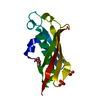
| |||||||||
|---|---|---|---|---|---|---|---|---|---|---|
| 1 | 
| |||||||||
| Unit cell |
| |||||||||
| Components on special symmetry positions |
|
-Components
| #1: Protein | Mass: 15416.212 Da / Num. of mol.: 1 Source method: isolated from a genetically manipulated source Source: (gene. exp.) Streptomyces sahachiroi (bacteria) / Gene: azi13 / Plasmid: pET24 / Production host: | ||
|---|---|---|---|
| #2: Chemical | ChemComp-5NE /   Mass: 186.207 Da / Num. of mol.: 1 / Source method: obtained synthetically / Formula: C12H10O2 Mass: 186.207 Da / Num. of mol.: 1 / Source method: obtained synthetically / Formula: C12H10O2 | ||
| #3: Chemical |   Mass: 96.063 Da / Num. of mol.: 2 / Source method: isolated from a natural source / Formula: SO4 Mass: 96.063 Da / Num. of mol.: 2 / Source method: isolated from a natural source / Formula: SO4#4: Water | ChemComp-HOH / |  Mass: 18.015 Da / Num. of mol.: 30 / Source method: isolated from a natural source / Formula: H2O Mass: 18.015 Da / Num. of mol.: 30 / Source method: isolated from a natural source / Formula: H2O |
-Experimental details
-Experiment
| Experiment | Method: X-RAY DIFFRACTION / Number of used crystals: 1 |
|---|
- Sample preparation
| Crystal | Density Matthews: 2.61 Å3/Da / Density % sol: 52.91 % |
|---|---|
| Crystal grow | Temperature: 298 K / Method: vapor diffusion, hanging drop / pH: 6 Details: 1.25 M ammonium sulfate and 0.1 M cacodylate pH 6.0 |
-Data collection
| Diffraction | Mean temperature: 100 K | ||||||||||||||||||
|---|---|---|---|---|---|---|---|---|---|---|---|---|---|---|---|---|---|---|---|
| Diffraction source | Source: SYNCHROTRON / Site: APS  / Beamline: 24-ID-C / Wavelength: 0.9792 Å / Beamline: 24-ID-C / Wavelength: 0.9792 Å | ||||||||||||||||||
| Detector | Type: DECTRIS PILATUS 6M-F / Detector: PIXEL / Date: Feb 12, 2015 | ||||||||||||||||||
| Radiation | Protocol: SINGLE WAVELENGTH / Monochromatic (M) / Laue (L): M / Scattering type: x-ray | ||||||||||||||||||
| Radiation wavelength | Wavelength: 0.9792 Å / Relative weight: 1 | ||||||||||||||||||
| Reflection | Resolution: 1.95→70.14 Å / Num. obs: 12265 / % possible obs: 99.4 % / Redundancy: 7 % / Biso Wilson estimate: 44.15 Å2 / CC1/2: 1 / Rmerge(I) obs: 0.056 / Rpim(I) all: 0.023 / Net I/σ(I): 20.9 / Num. measured all: 85401 | ||||||||||||||||||
| Reflection shell |
|
- Processing
| Software |
| ||||||||||||||||||||||||||||
|---|---|---|---|---|---|---|---|---|---|---|---|---|---|---|---|---|---|---|---|---|---|---|---|---|---|---|---|---|---|
| Refinement | Method to determine structure: FOURIER SYNTHESIS Starting model: D_1000217232 Resolution: 2.2→61.023 Å / SU ML: 0.24 / Cross valid method: FREE R-VALUE / σ(F): 1.38 / Phase error: 26.96
| ||||||||||||||||||||||||||||
| Solvent computation | Shrinkage radii: 0.9 Å / VDW probe radii: 1.11 Å | ||||||||||||||||||||||||||||
| Displacement parameters | Biso max: 94.36 Å2 / Biso mean: 48.3142 Å2 / Biso min: 32.81 Å2 | ||||||||||||||||||||||||||||
| Refinement step | Cycle: final / Resolution: 2.2→61.023 Å
| ||||||||||||||||||||||||||||
| Refine LS restraints |
| ||||||||||||||||||||||||||||
| LS refinement shell | Refine-ID: X-RAY DIFFRACTION / Total num. of bins used: 3
|
