 Movie
Movie Controller

[English] 日本語
 Yorodumi
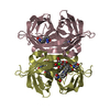
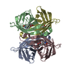
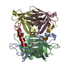
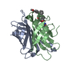
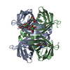
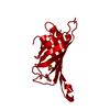
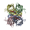
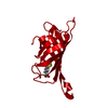
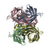
- PDB-5n8j: CRYSTAL STRUCTURE OF STREPTAVIDIN WITH PEPTIDE D-amino acid conta... -
+ Open data

- Basic information
| Entry | Database: PDB / ID: 5n8j | ||||||
|---|---|---|---|---|---|---|---|
| Title | CRYSTAL STRUCTURE OF STREPTAVIDIN WITH PEPTIDE D-amino acid containing peptide GyGlanvdessG | ||||||
 Components Components |
| ||||||
 Keywords Keywords | STREPTAVIDIN PEPTIDE 11101 COMPLEX / STREPTAVIDIN / BIOTIN BINDING / D-AMINO ACIDS | ||||||
| Function / homology |  Function and homology information Function and homology information | ||||||
| Biological species |  Streptomyces avidinii (bacteria) Streptomyces avidinii (bacteria)synthetic construct (others) | ||||||
| Method |  X-RAY DIFFRACTION / SYNCHROTRON / MOLECULAR REPLACEMENT / Resolution: 1.05 Å X-RAY DIFFRACTION / SYNCHROTRON / MOLECULAR REPLACEMENT / Resolution: 1.05 Å | ||||||
 Authors Authors | Lyamichev, V. / Goodrich, L. / Sullivan, E. / Bannen, R. / Benz, J. / Albert, T. / Patel, J. | ||||||
 Citation Citation | Journal: Sci Rep / Year: 2017 Title: Stepwise Evolution Improves Identification of Diverse Peptides Binding to a Protein Target. Authors: Lyamichev, V.I. / Goodrich, L.E. / Sullivan, E.H. / Bannen, R.M. / Benz, J. / Albert, T.J. / Patel, J.J. | ||||||
| History |
|
- Structure visualization
| Structure viewer | Molecule: MolmilJmol/JSmol |
|---|
- Downloads & links
-Download
| PDBx/mmCIF format | 5n8j.cif.gz | 238.6 KB | Display | PDBx/mmCIF format |
|---|---|---|---|---|
| PDB format | pdb5n8j.ent.gz | 190.1 KB | Display | PDB format |
| PDBx/mmJSON format | 5n8j.json.gz | Tree view | PDBx/mmJSON format | |
| Others |  Other downloads Other downloads |
-Validation report
| Arichive directory | https://data.pdbj.org/pub/pdb/validation_reports/n8/5n8jftp://data.pdbj.org/pub/pdb/validation_reports/n8/5n8j | HTTPS FTP |
|---|
-Related structure data
| Related structure data |  5n7xC  5n89C  5n8bC  5n8eC  5n8tC 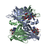 5n8wC  5n99C C: citing same article ( |
|---|---|
| Similar structure data |

-Links
 PDBj
PDBj
- Assembly
| Deposited unit | 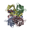
| ||||||||
|---|---|---|---|---|---|---|---|---|---|
| 1 |
| ||||||||
| Unit cell |
|
-Components
| #1: Protein | Mass: 18849.672 Da / Num. of mol.: 4 Source method: isolated from a genetically manipulated source Source: (gene. exp.) Streptomyces avidinii (bacteria) / Production host: #2: Polypeptide(D) | Mass: 1168.171 Da / Num. of mol.: 3 / Source method: obtained synthetically / Source: (synth.) synthetic construct (others) #3: Chemical | ChemComp-IPA / |   Mass: 60.095 Da / Num. of mol.: 1 / Source method: obtained synthetically / Formula: C3H8O / Comment: alkaloid*YM Mass: 60.095 Da / Num. of mol.: 1 / Source method: obtained synthetically / Formula: C3H8O / Comment: alkaloid*YM#4: Water | ChemComp-HOH / |  Mass: 18.015 Da / Num. of mol.: 689 / Source method: isolated from a natural source / Formula: H2O Mass: 18.015 Da / Num. of mol.: 689 / Source method: isolated from a natural source / Formula: H2OHas protein modification | N | |
|---|
-Experimental details
-Experiment
| Experiment | Method: X-RAY DIFFRACTION / Number of used crystals: 1 |
|---|
- Sample preparation
| Crystal grow | Temperature: 293 K / Method: vapor diffusion, sitting drop Details: 0.1M Sodium citrate pH 5.6 20% PEG4000 20% Isopropanol |
|---|
-Data collection
| Diffraction | Mean temperature: 100 K |
|---|---|
| Diffraction source | Source: SYNCHROTRON / Site: SLS  / Beamline: X10SA / Wavelength: 0.700002 Å / Beamline: X10SA / Wavelength: 0.700002 Å |
| Detector | Type: DECTRIS PILATUS 6M-F / Detector: PIXEL / Date: Sep 22, 2015 |
| Radiation | Protocol: SINGLE WAVELENGTH / Monochromatic (M) / Laue (L): M / Scattering type: x-ray |
| Radiation wavelength | Wavelength: 0.700002 Å / Relative weight: 1 |
| Reflection | Resolution: 1.05→57.09 Å / Num. obs: 195681 / % possible obs: 99.6 % / Redundancy: 3.43 % / CC1/2: 0.999 / Rsym value: 0.036 / Net I/σ(I): 11.4 |
| Reflection shell | Resolution: 1.05→1.14 Å / Redundancy: 3.4 % / Mean I/σ(I) obs: 1.11 / Num. unique obs: 44813 / CC1/2: 0.388 / Rsym value: 0.64 / % possible all: 99.7 |
- Processing
| Software |
| ||||||||||||||||||||||||||||||||||||||||||||||||||||||||||||||||||||||||||||||||||||||||||||||||||||||||||||||||||||||||||||||||||||||||||||||||||||||||||||||||||||||||||||||||||||||
|---|---|---|---|---|---|---|---|---|---|---|---|---|---|---|---|---|---|---|---|---|---|---|---|---|---|---|---|---|---|---|---|---|---|---|---|---|---|---|---|---|---|---|---|---|---|---|---|---|---|---|---|---|---|---|---|---|---|---|---|---|---|---|---|---|---|---|---|---|---|---|---|---|---|---|---|---|---|---|---|---|---|---|---|---|---|---|---|---|---|---|---|---|---|---|---|---|---|---|---|---|---|---|---|---|---|---|---|---|---|---|---|---|---|---|---|---|---|---|---|---|---|---|---|---|---|---|---|---|---|---|---|---|---|---|---|---|---|---|---|---|---|---|---|---|---|---|---|---|---|---|---|---|---|---|---|---|---|---|---|---|---|---|---|---|---|---|---|---|---|---|---|---|---|---|---|---|---|---|---|---|---|---|---|
| Refinement | Method to determine structure: MOLECULAR REPLACEMENT / Resolution: 1.05→57.09 Å / Cor.coef. Fo:Fc: 0.976 / Cor.coef. Fo:Fc free: 0.969 / SU B: 1.333 / SU ML: 0.028 / Cross valid method: THROUGHOUT / ESU R: 0.031 / ESU R Free: 0.032 / Details: HYDROGENS HAVE BEEN ADDED IN THE RIDING POSITIONS
| ||||||||||||||||||||||||||||||||||||||||||||||||||||||||||||||||||||||||||||||||||||||||||||||||||||||||||||||||||||||||||||||||||||||||||||||||||||||||||||||||||||||||||||||||||||||
| Solvent computation | Ion probe radii: 0.8 Å / Shrinkage radii: 0.8 Å / VDW probe radii: 1.2 Å | ||||||||||||||||||||||||||||||||||||||||||||||||||||||||||||||||||||||||||||||||||||||||||||||||||||||||||||||||||||||||||||||||||||||||||||||||||||||||||||||||||||||||||||||||||||||
| Displacement parameters | Biso mean: 16.321 Å2
| ||||||||||||||||||||||||||||||||||||||||||||||||||||||||||||||||||||||||||||||||||||||||||||||||||||||||||||||||||||||||||||||||||||||||||||||||||||||||||||||||||||||||||||||||||||||
| Refinement step | Cycle: 1 / Resolution: 1.05→57.09 Å
| ||||||||||||||||||||||||||||||||||||||||||||||||||||||||||||||||||||||||||||||||||||||||||||||||||||||||||||||||||||||||||||||||||||||||||||||||||||||||||||||||||||||||||||||||||||||
| Refine LS restraints |
|